Cranwich Pits

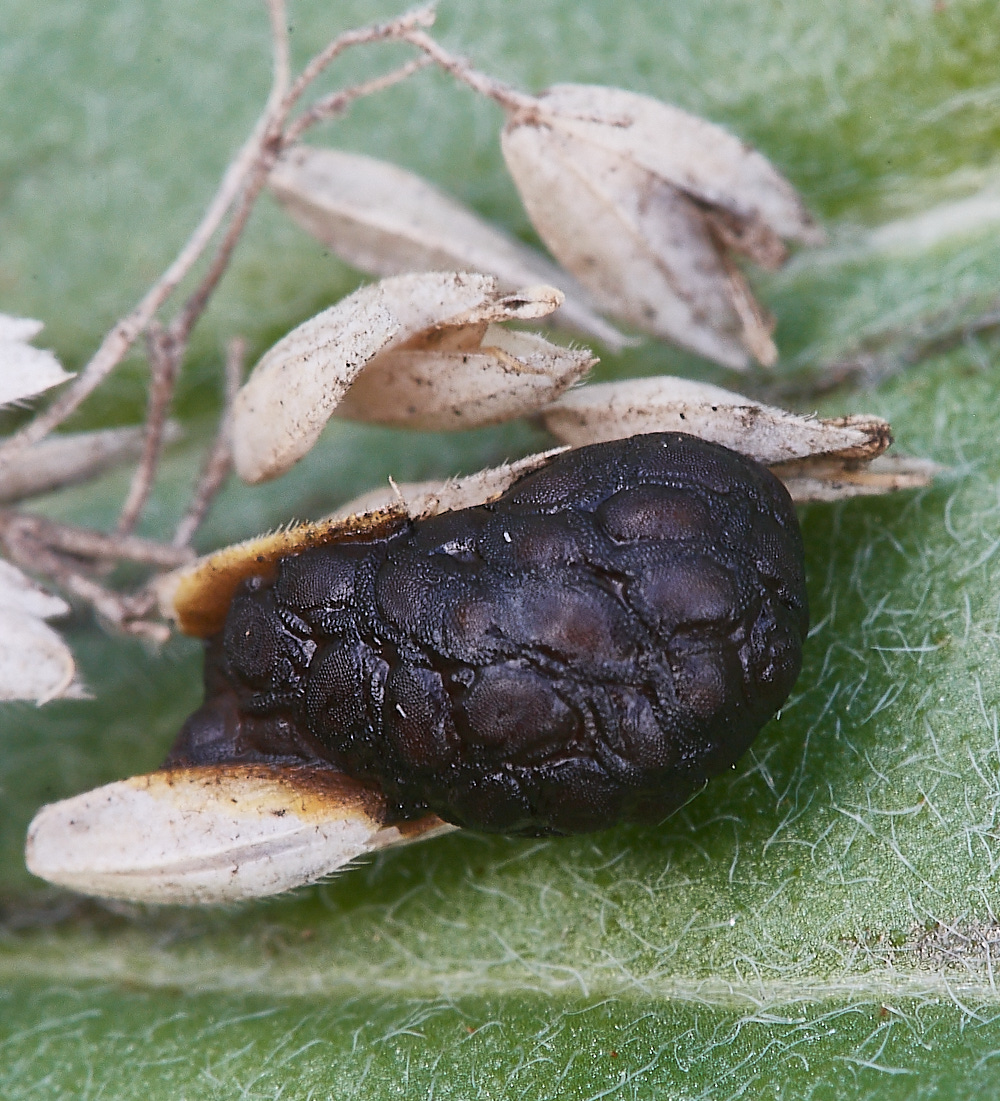
Something unusual with no one quite sure?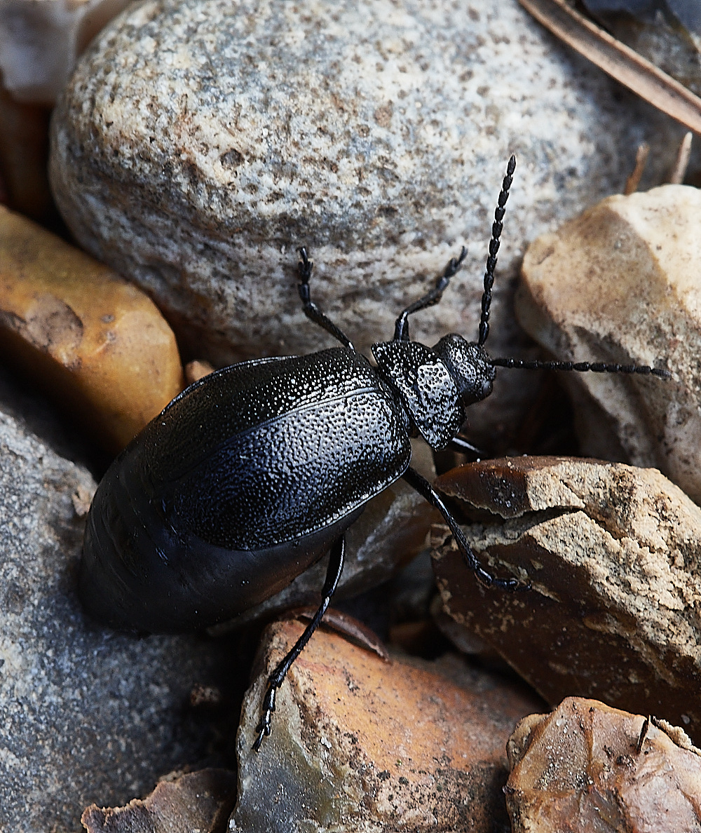
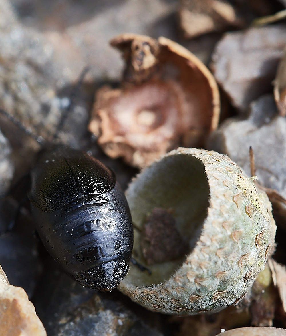
Beetle Sp ?
Clearly a pregnant female (Gravid) because the abdomen is bloated and it extends beyond the wing cases.
This turned out to Galeruca tanaceti and if so new for the site.
Thanks to Vanna & Tony for id.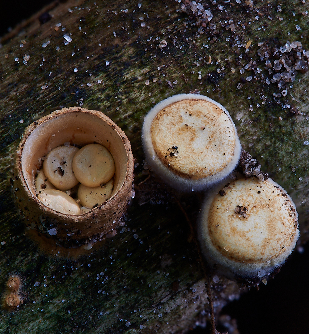
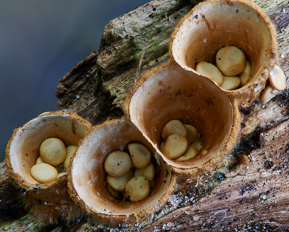
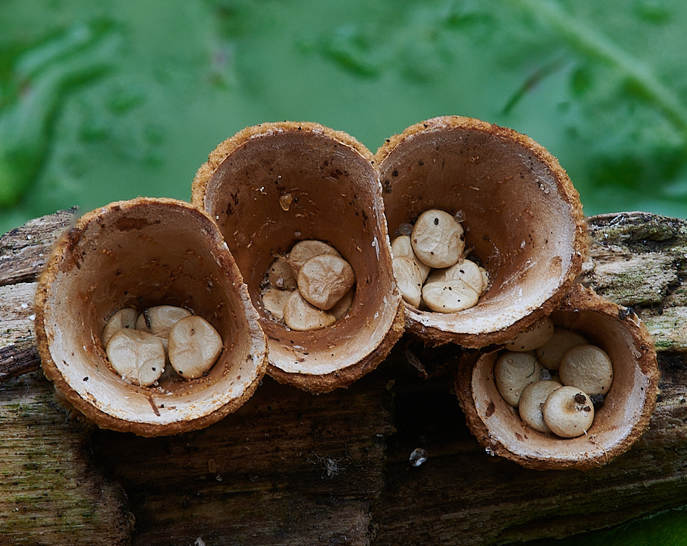
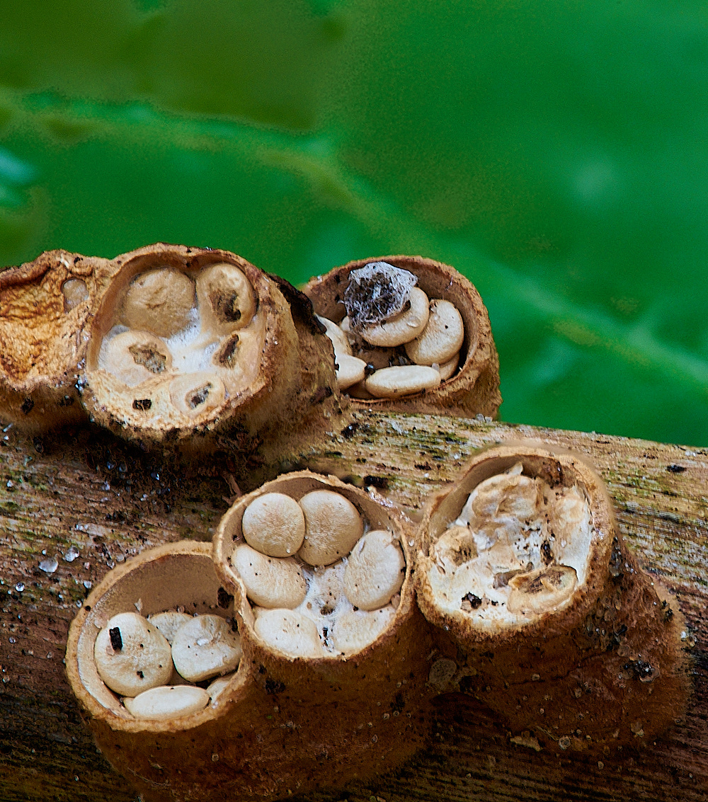
Common Bird's Nest Fungus (Crucibulum laeve)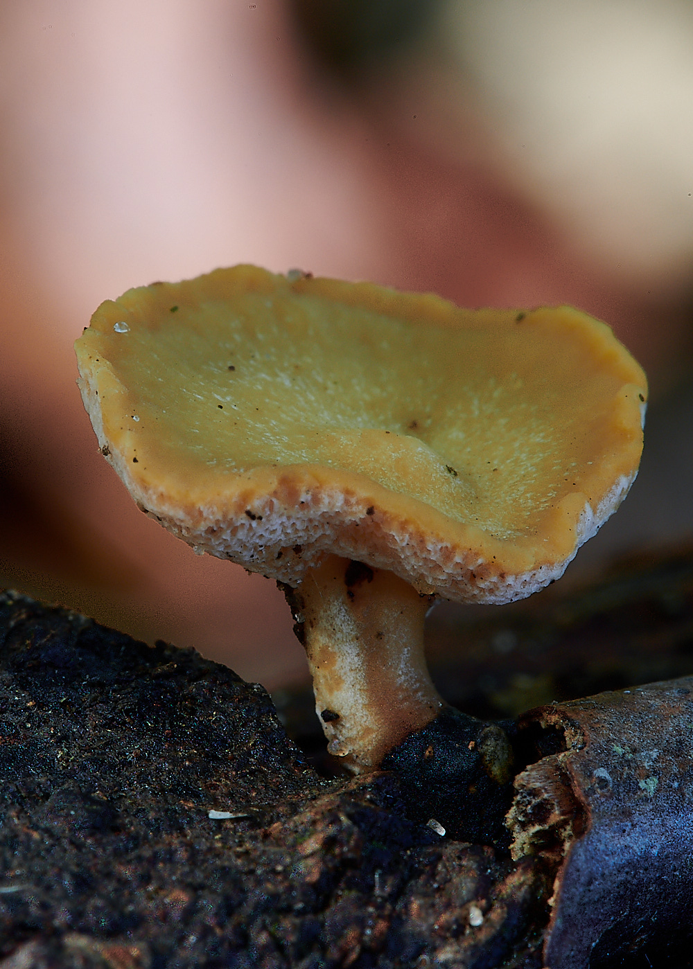
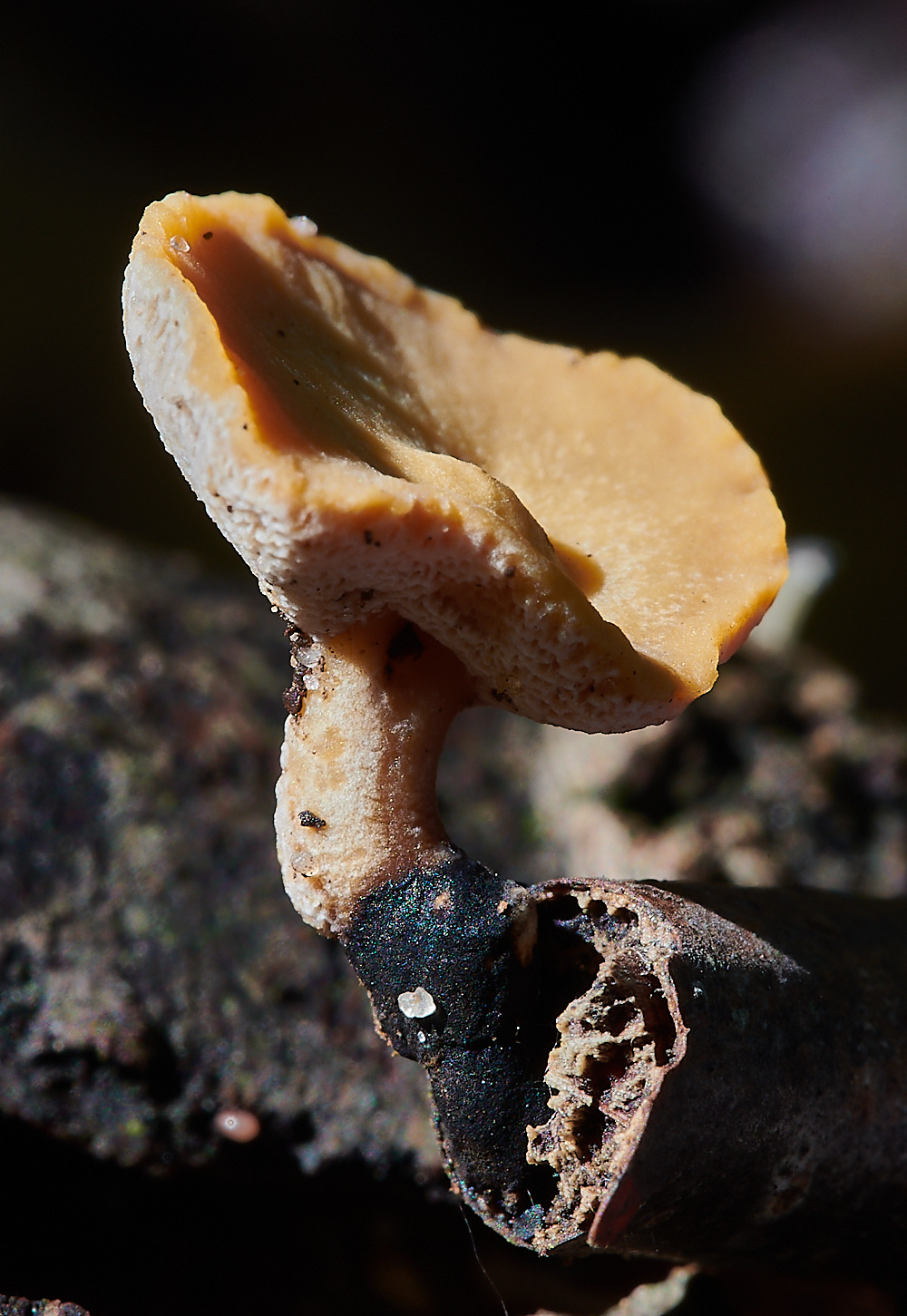
Blackfoot Polypore (Polyporus leptocephalus)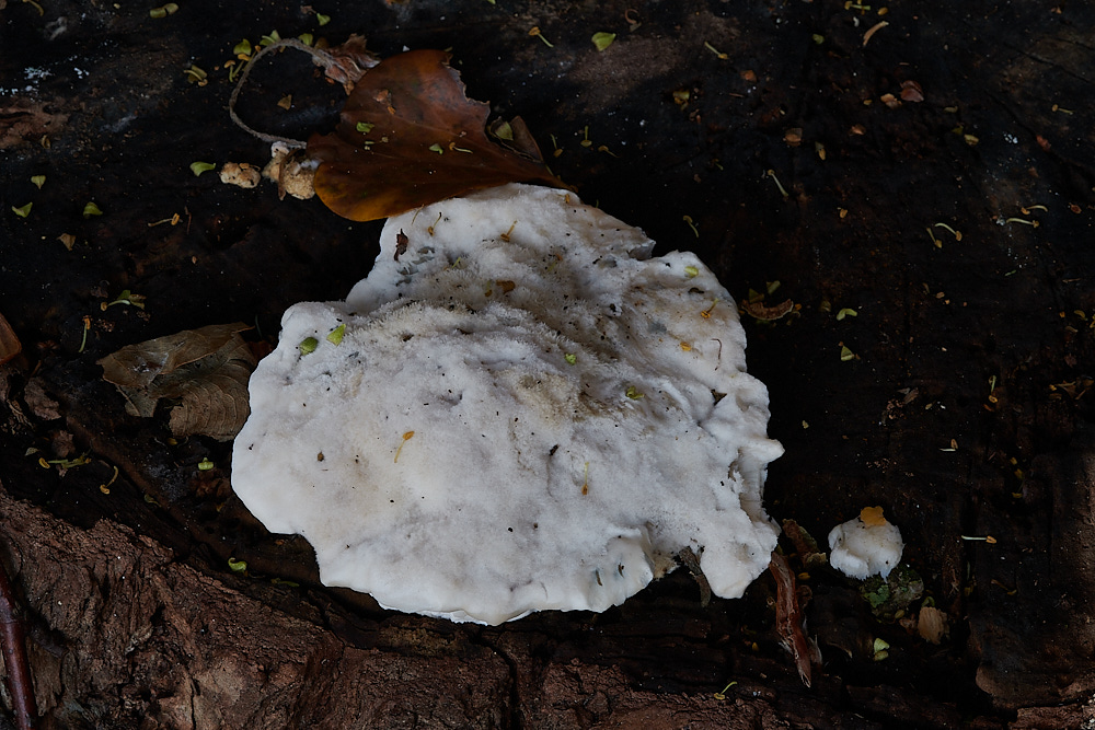
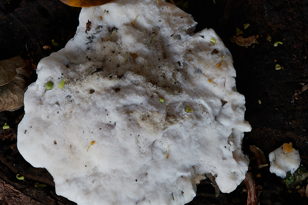
Blueing Bracket (Postia subcaesia)
Subtley different in form in that it is not semicircular.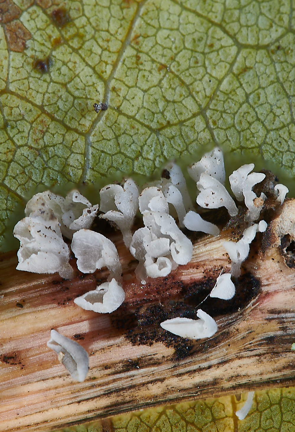
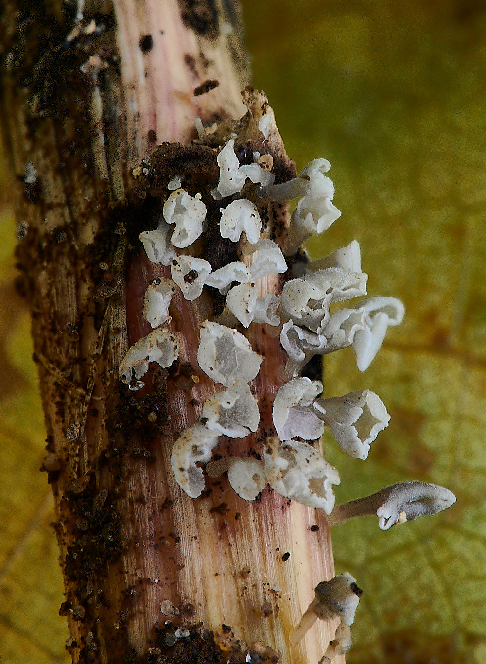
Calyptella capula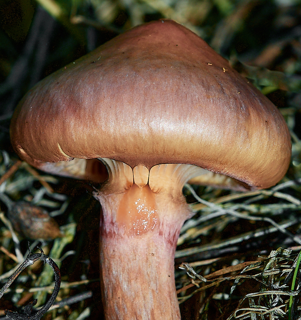
Copper Spike (Chromgomphus rutilus)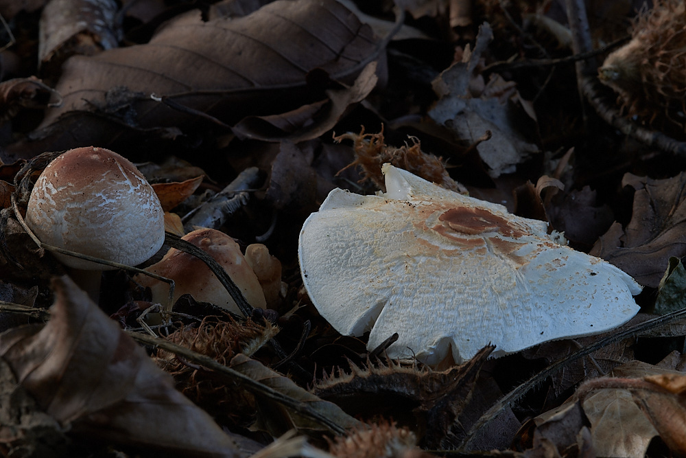
Dapperling Sp?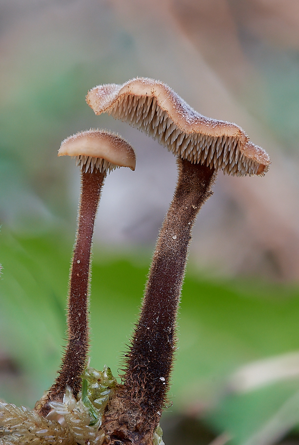
Earpick Fungus (Auricalpium vulgare) on a Pine Cone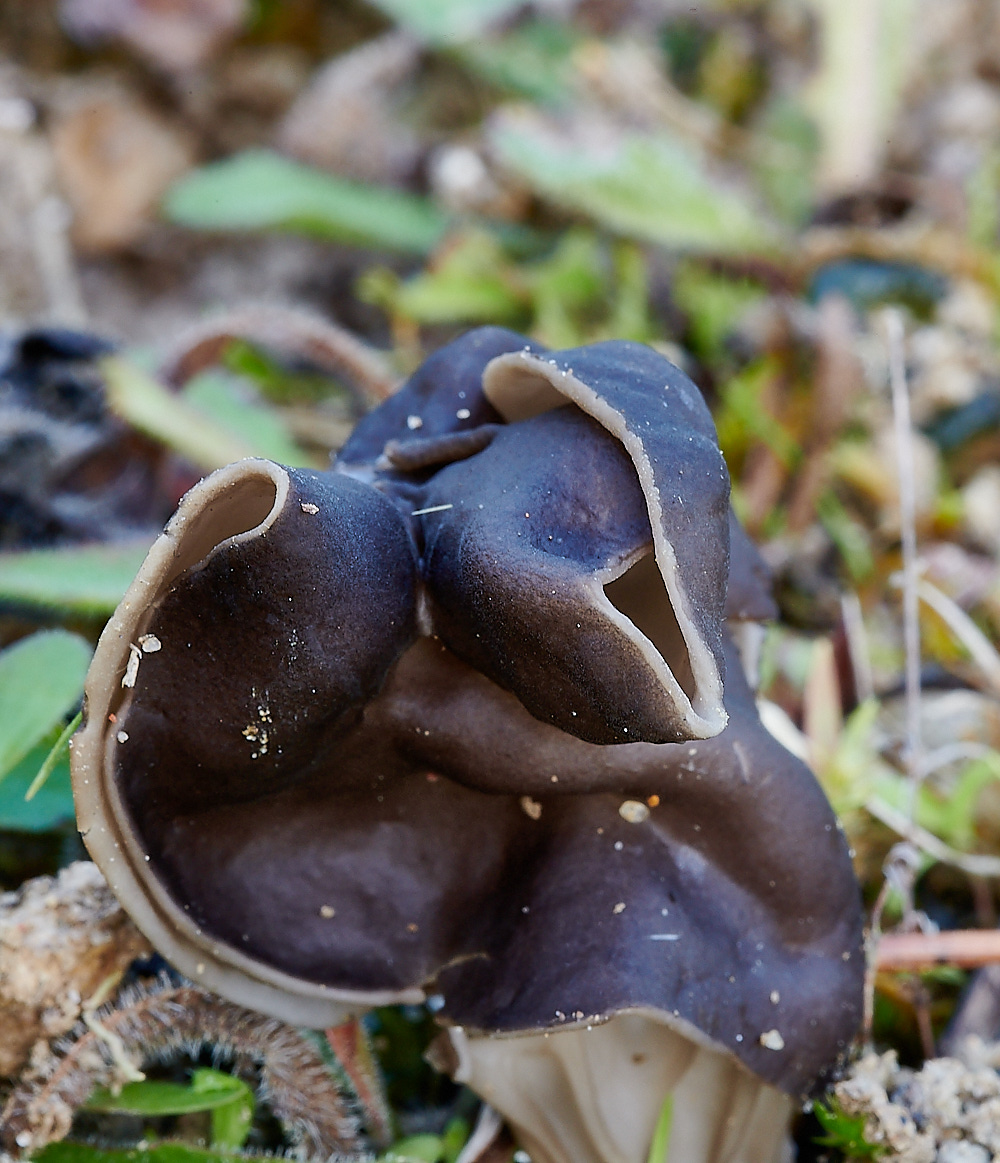
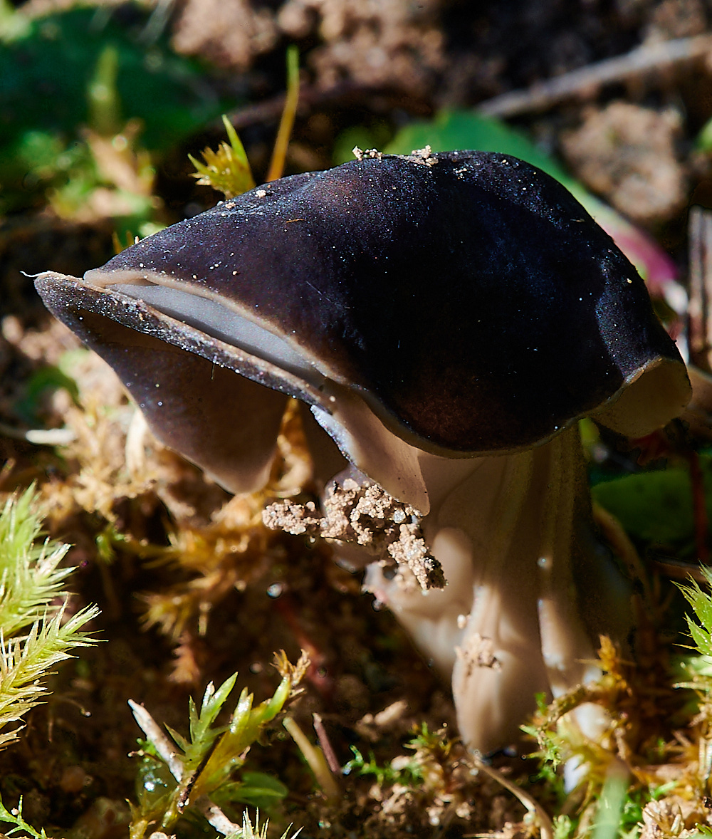
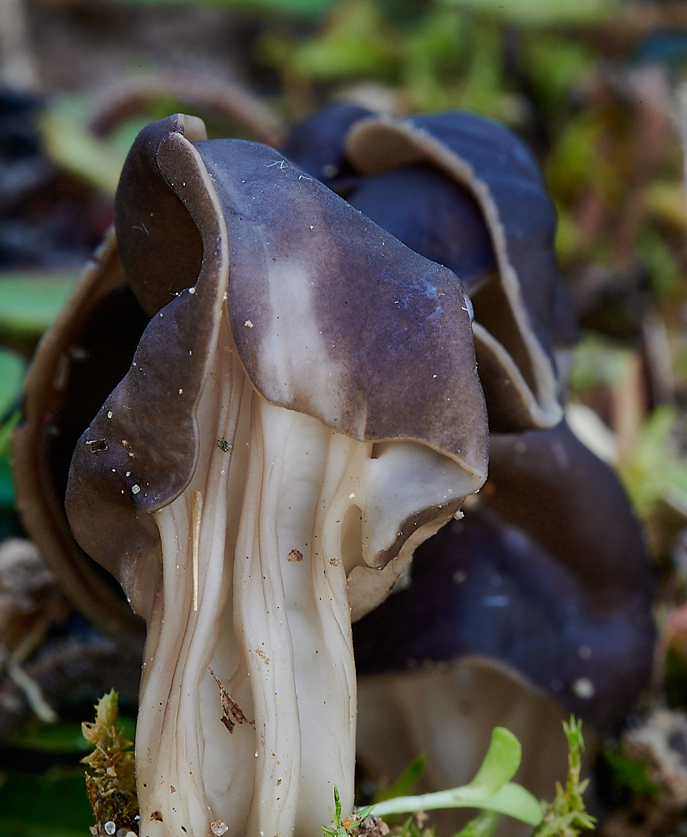
Elfin Saddle (Helvella lacunosa)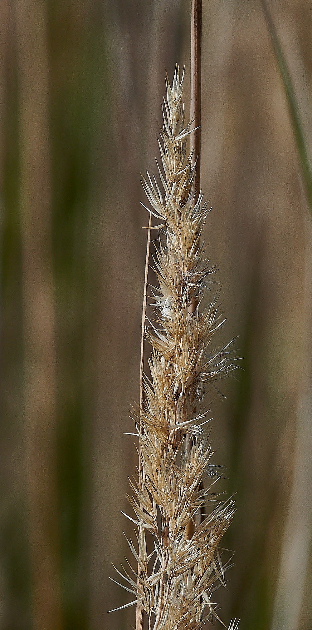
Calamagrostis Sp
Thanks to Vicky for id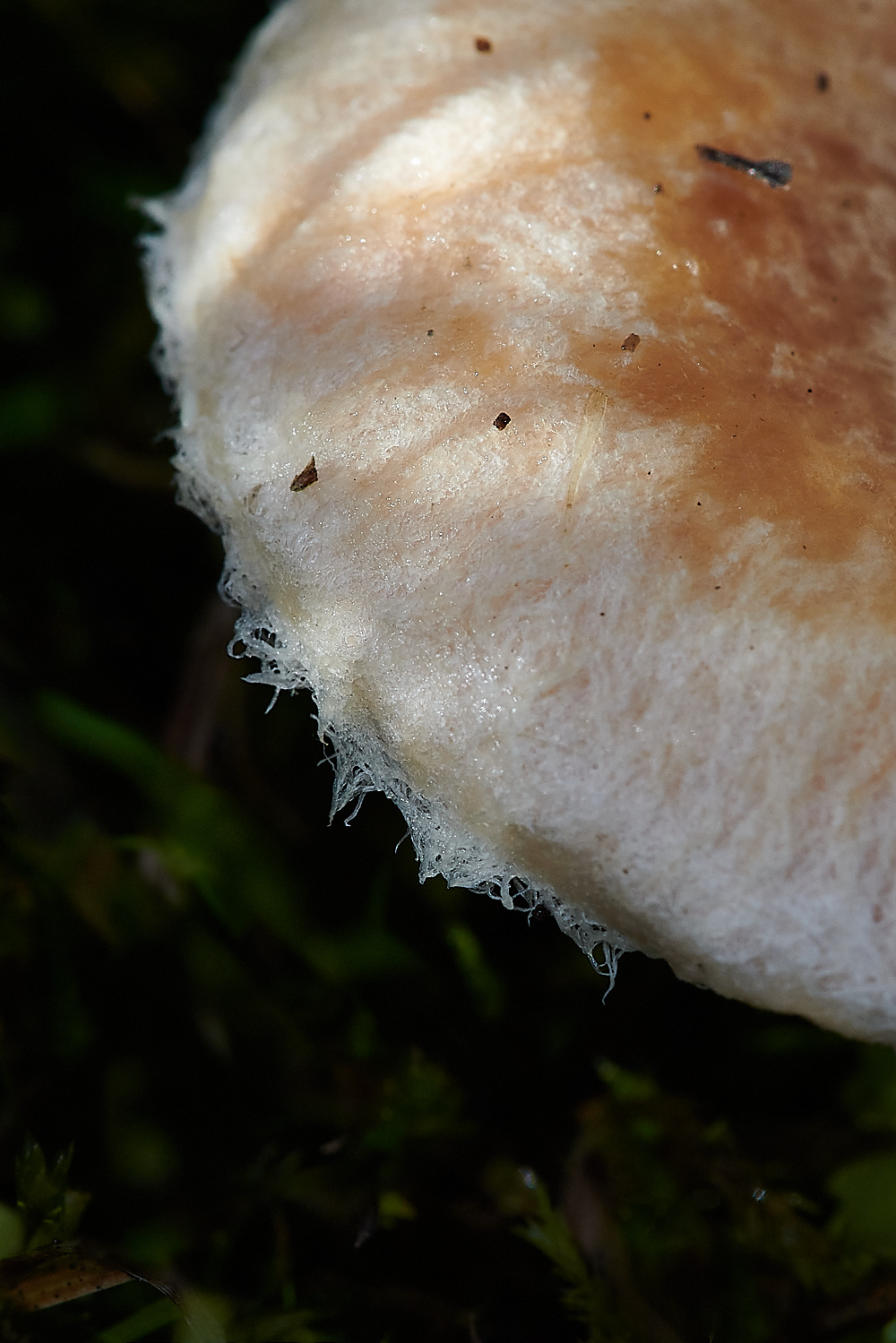
Bearded/Wooly Milkcap (Lactarius pubescens)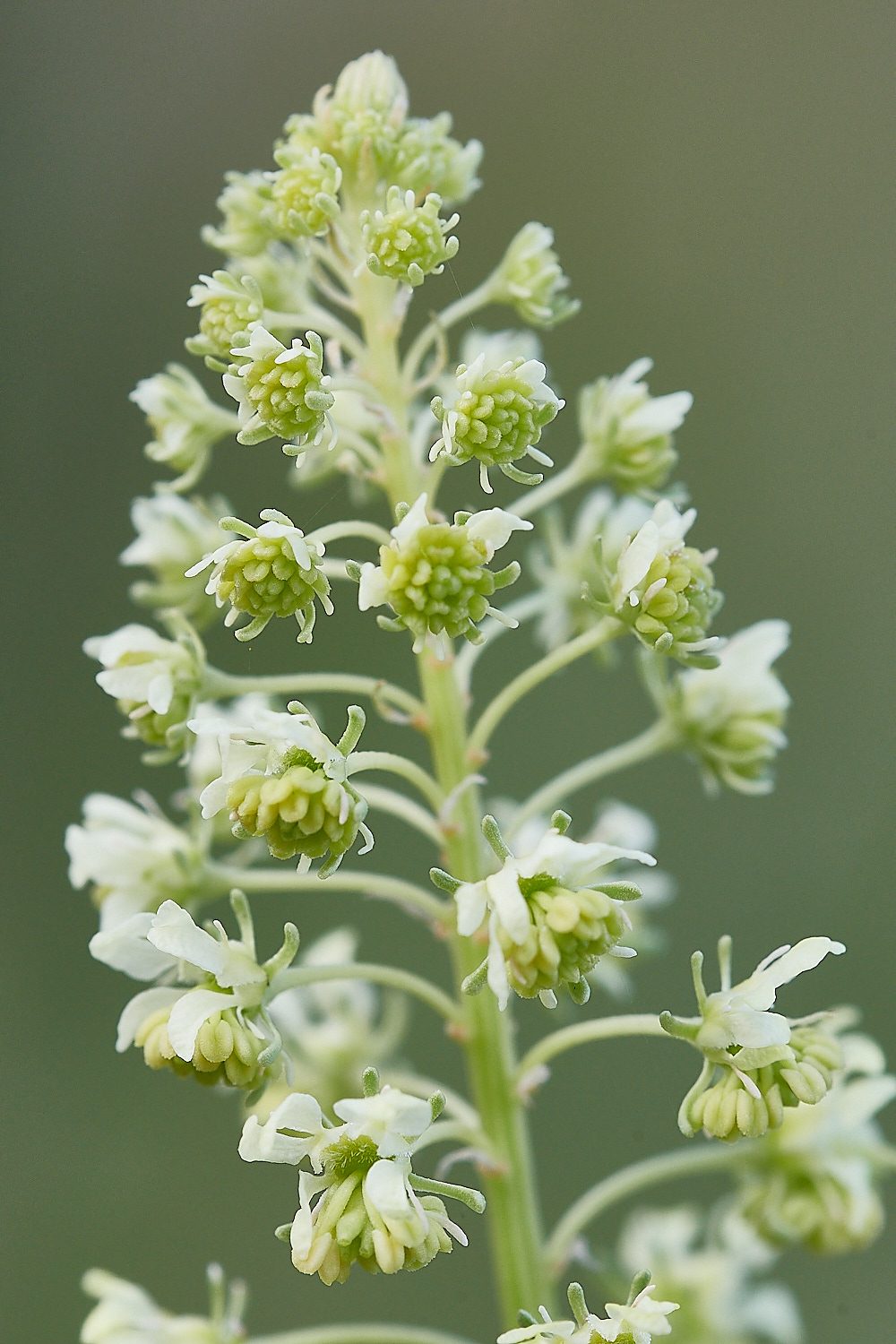
Wild Mignonette (Reseda lutea)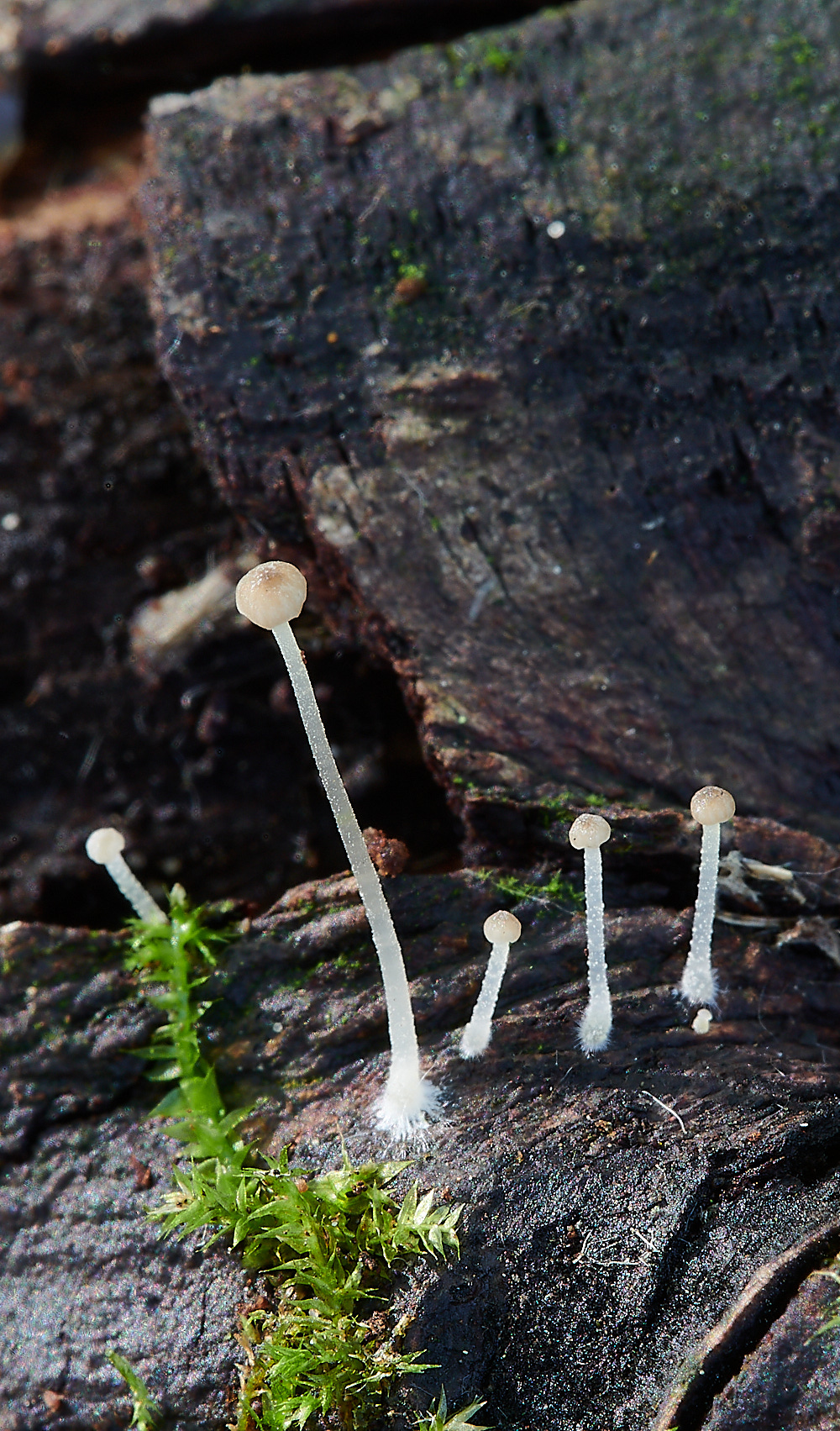
Mycena Sp?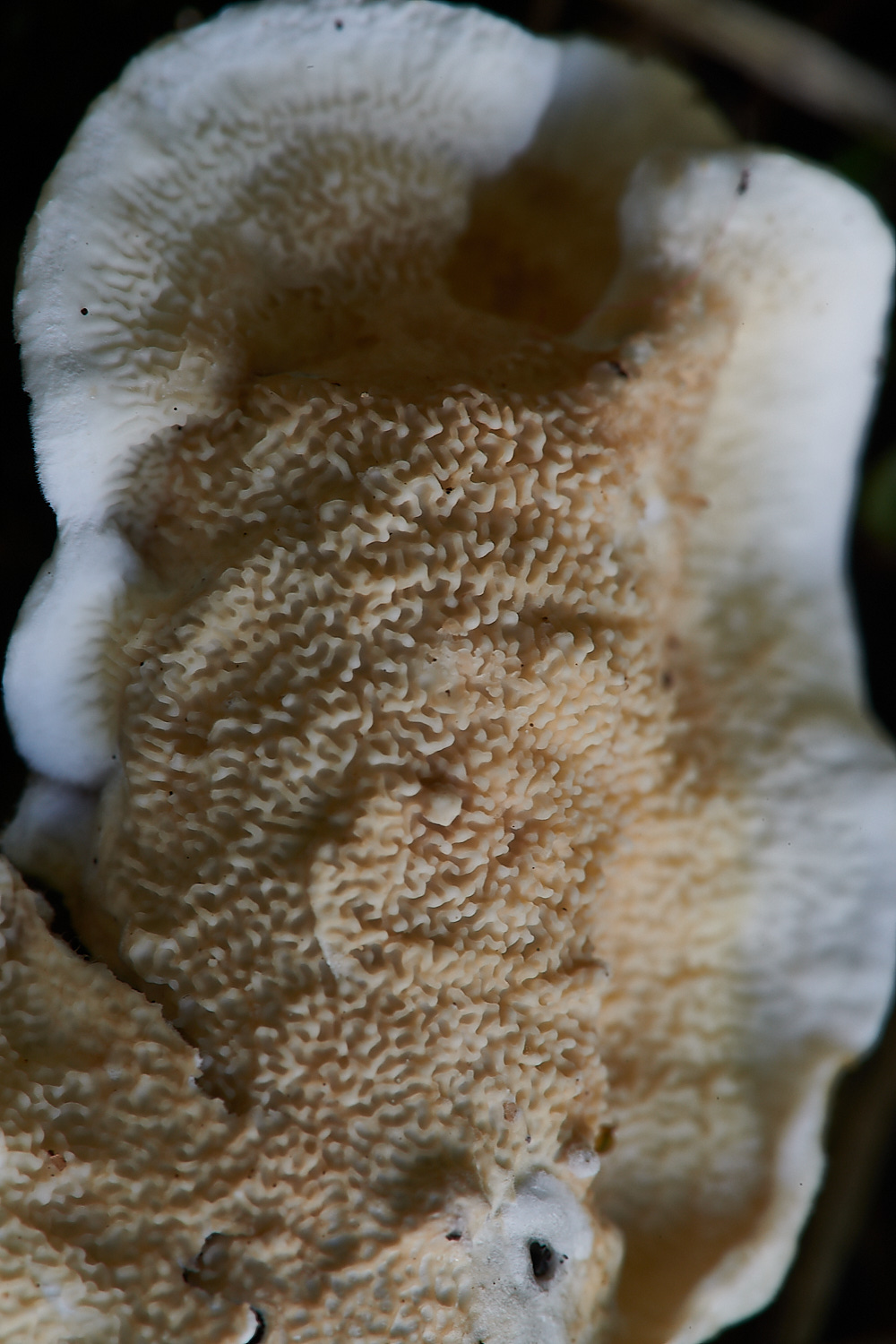
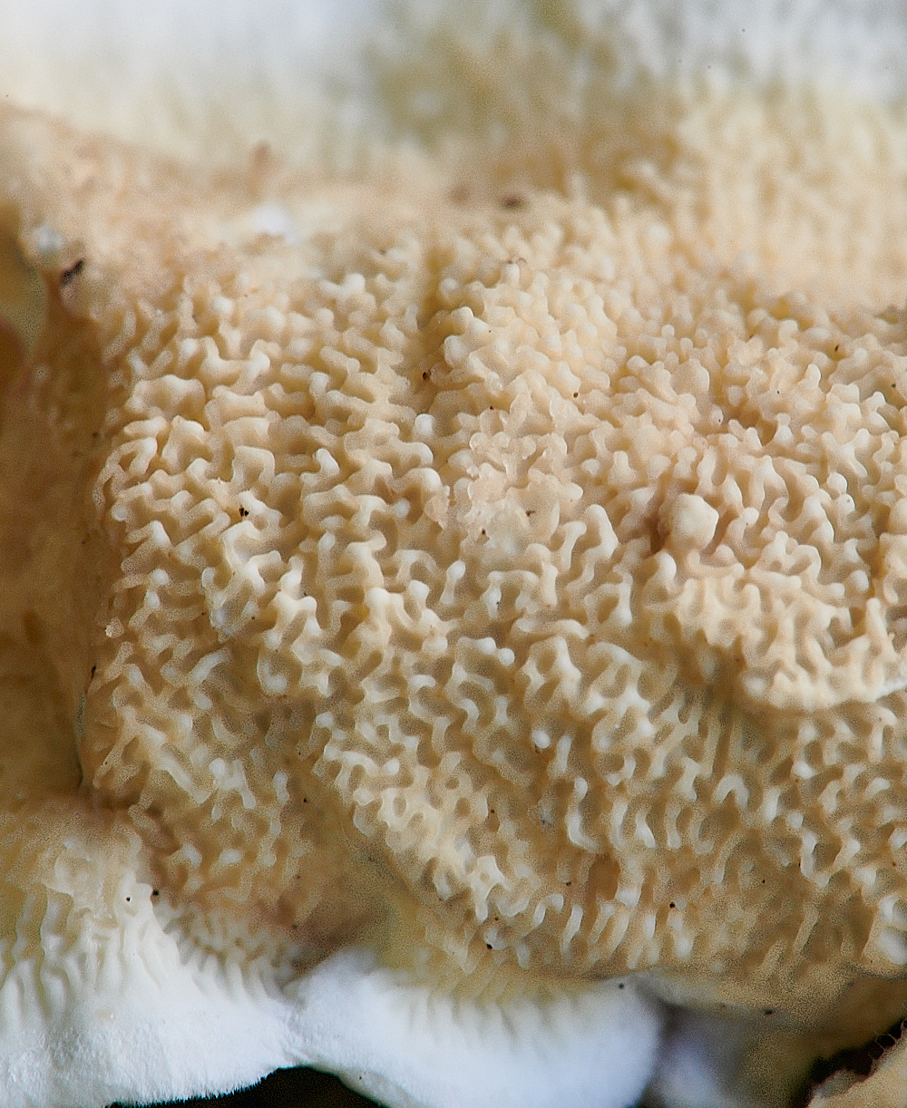
Netted Crust (Byssomerulius corium)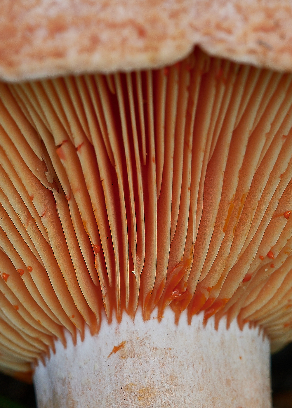
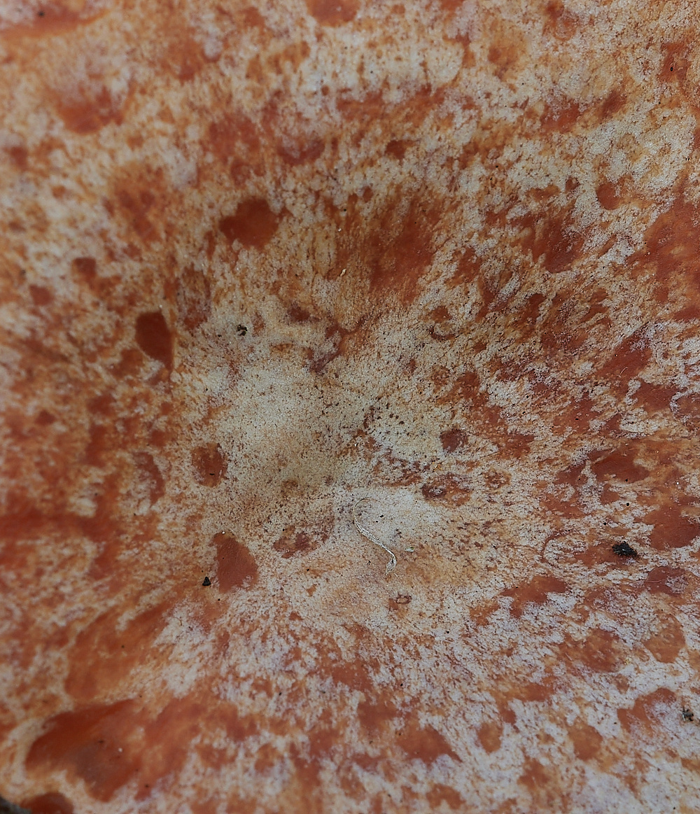
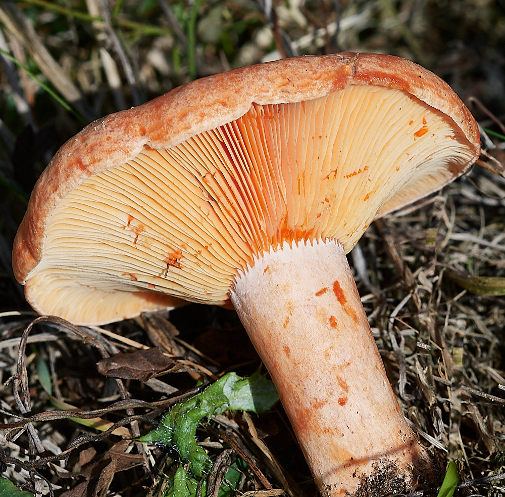
The stunning
Saffrom Milkcap (Lactarius deliciosus)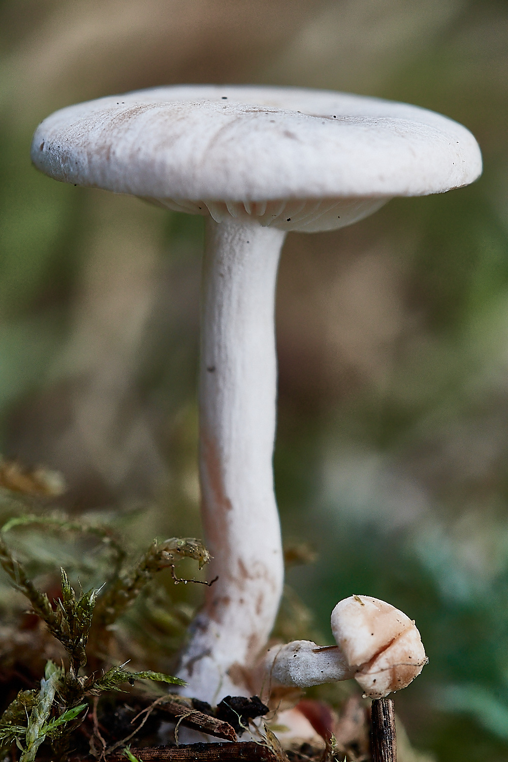
Fool's funnel (Clitocybe rivulosa)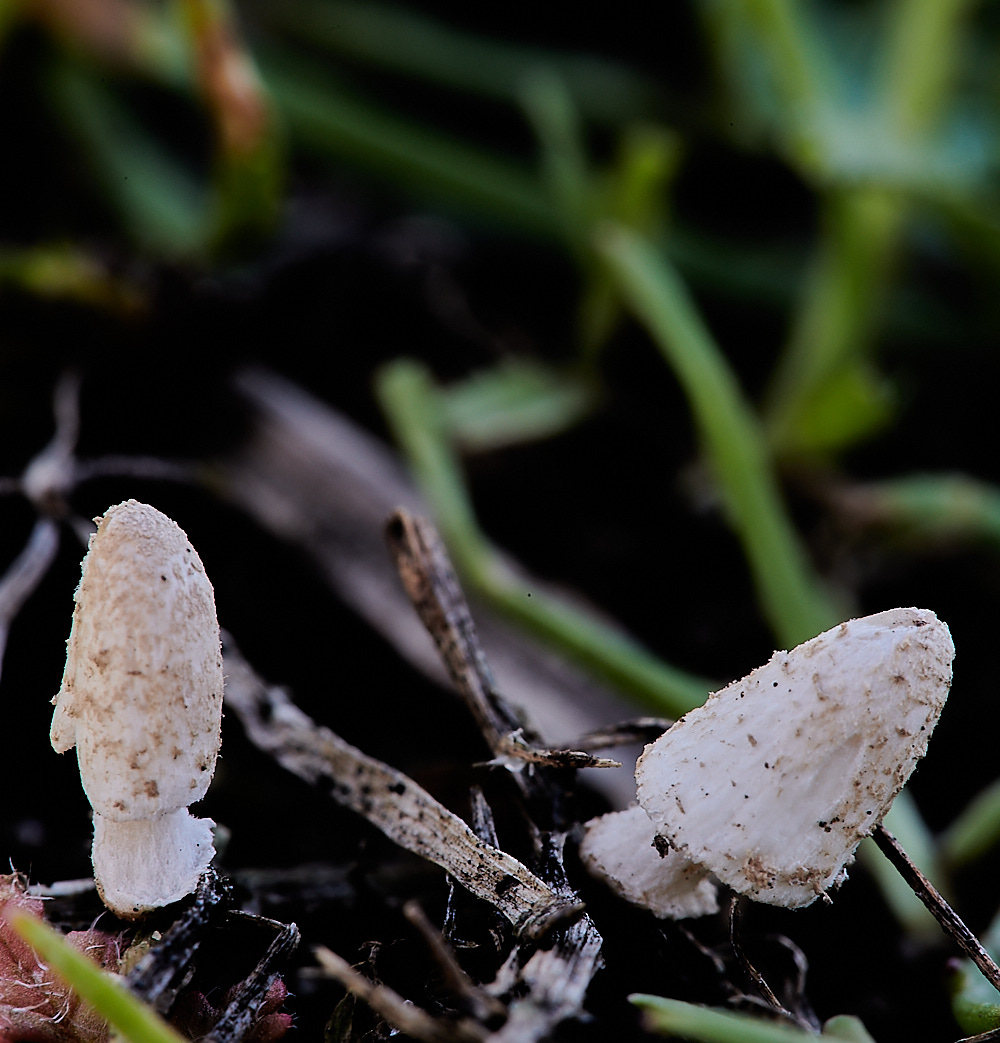
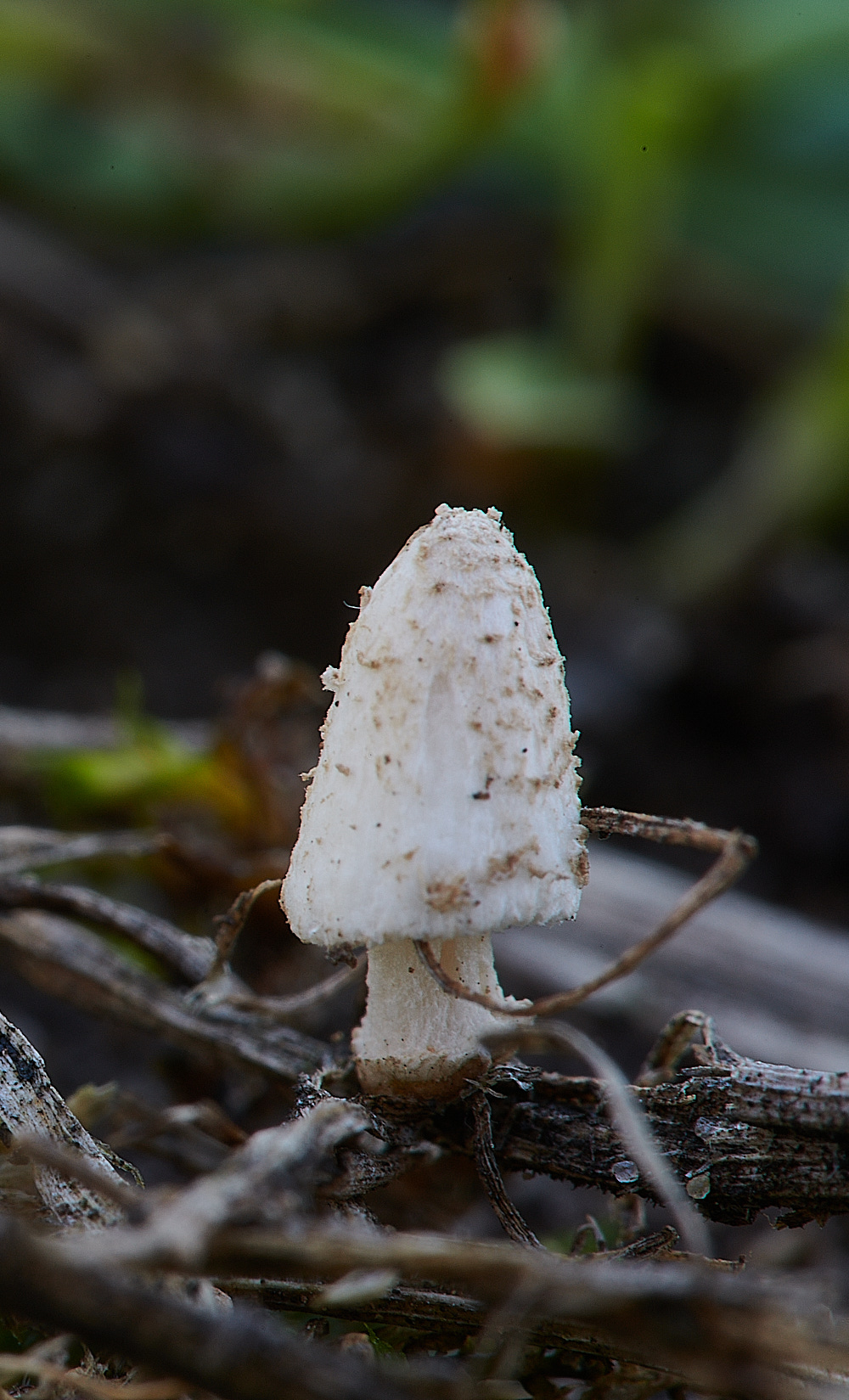
Something tiny and unknown. Awaiting microscopy. 3mm tall.
Yvonne's comments - I think this is a species of inkcap. I have had ones like this before and think they are sterile specimens.
They don't look particularly dry but they do look mature.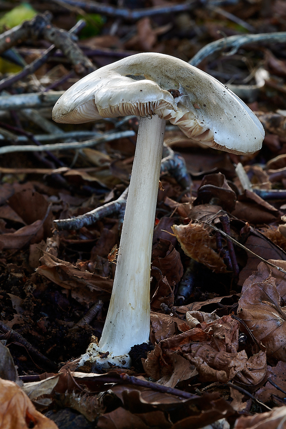
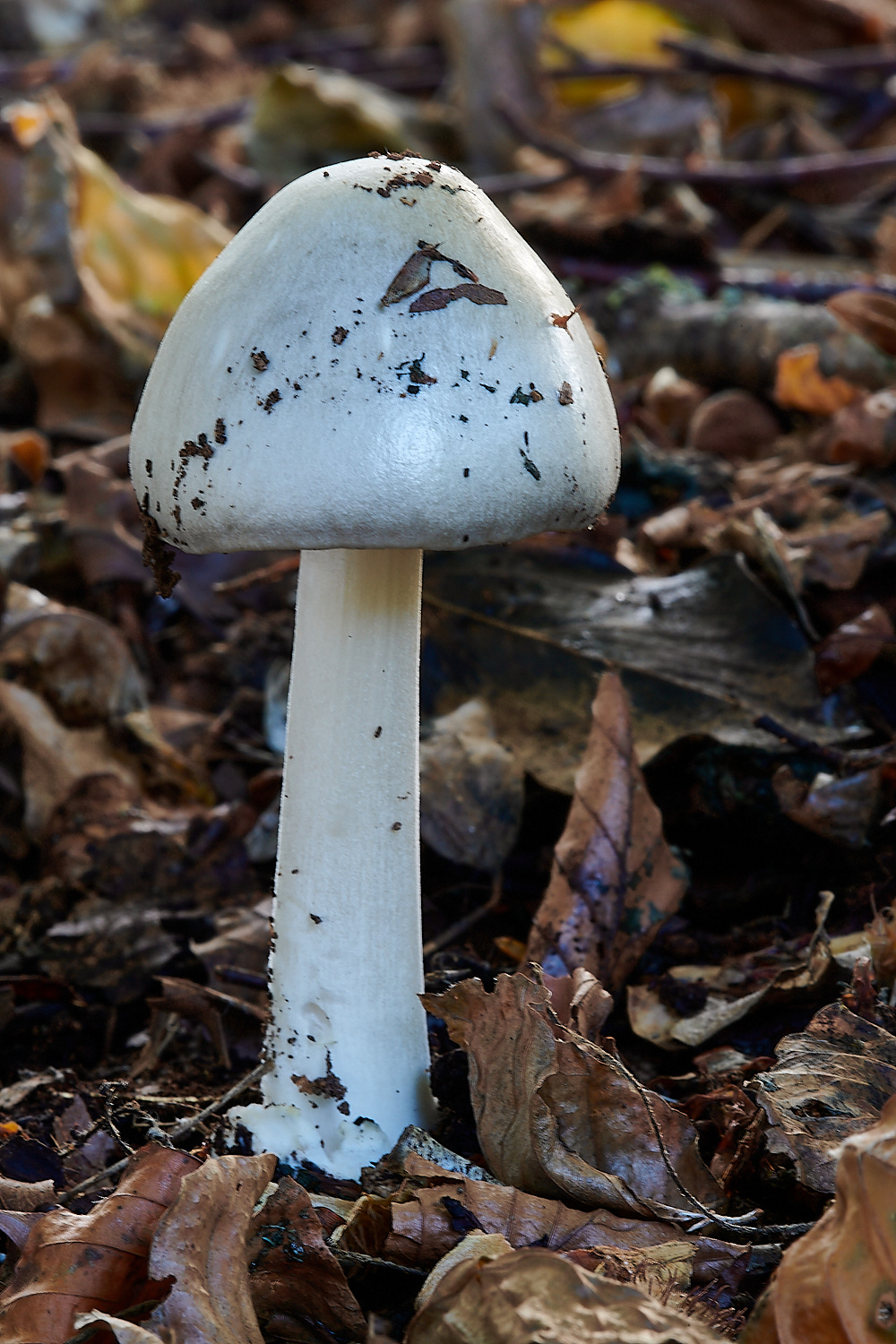
A very statuesque fungi
Stubble Rosegill (Volvopluteus gloiocephalus)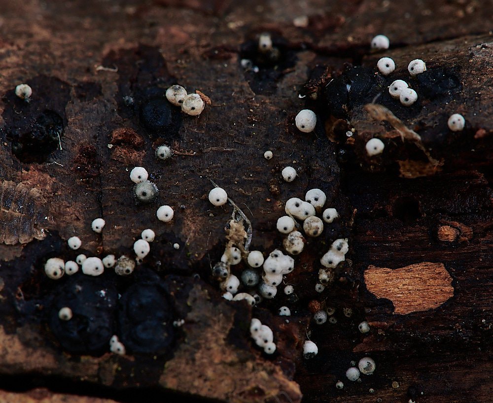
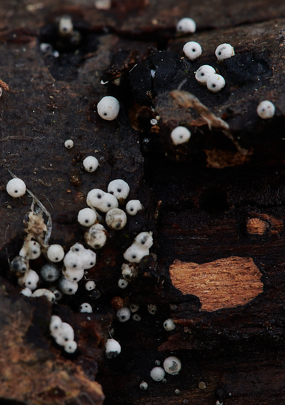
Woolly Woodwart (Lasiospheria ovina)
Barnham Broom Fen
Something from Hoveton to wet everyone's appetite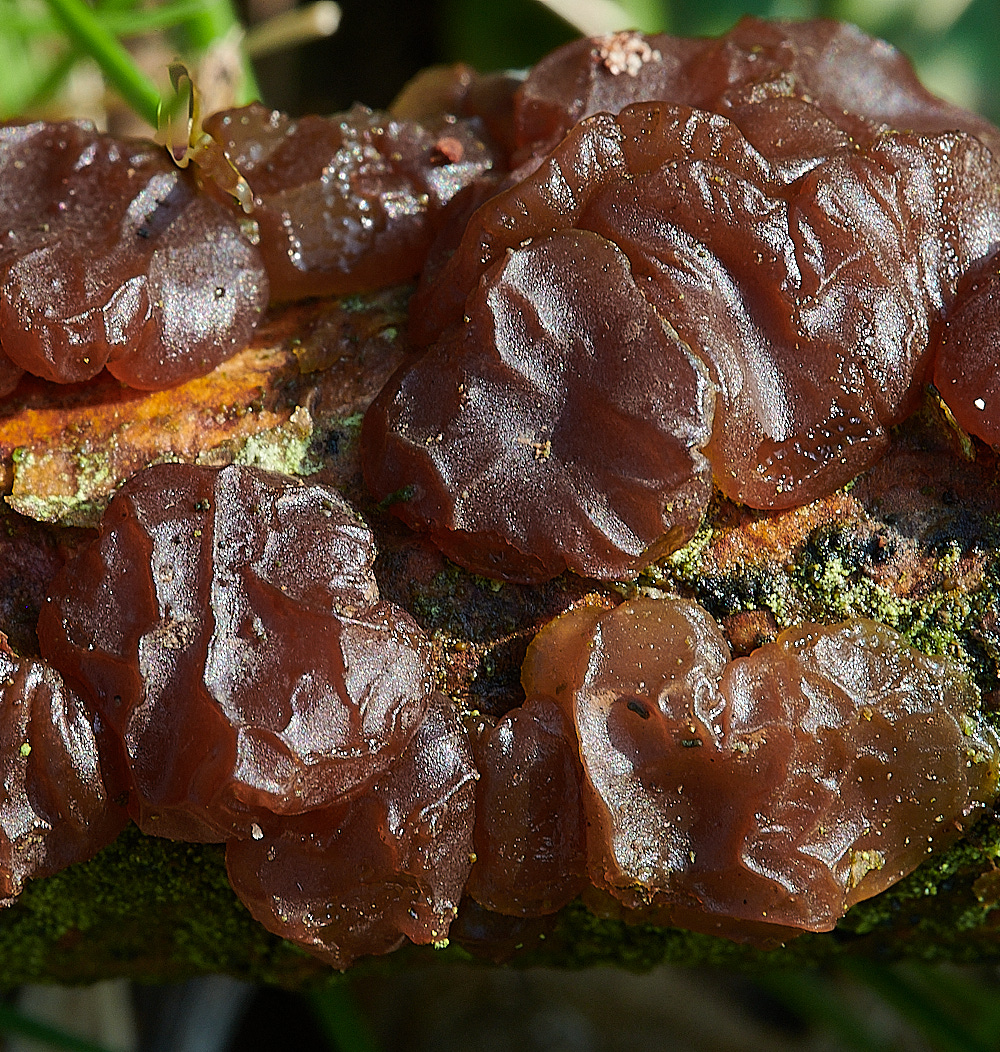
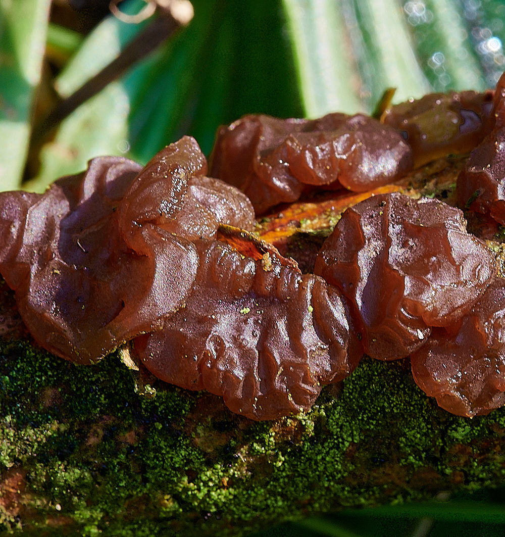
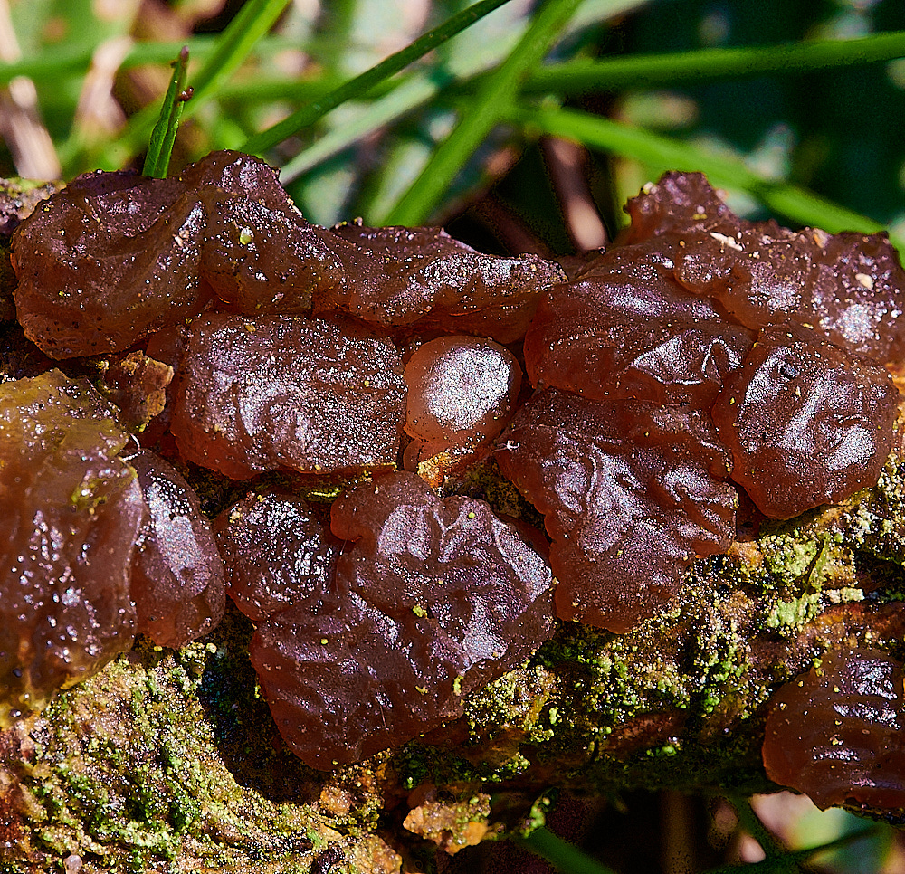
Sultanas on a stick?
Pine Jelly (Exidia saccharina)
Barnham Broom Fen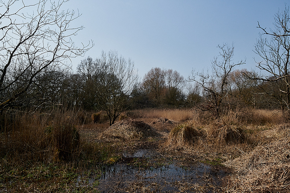
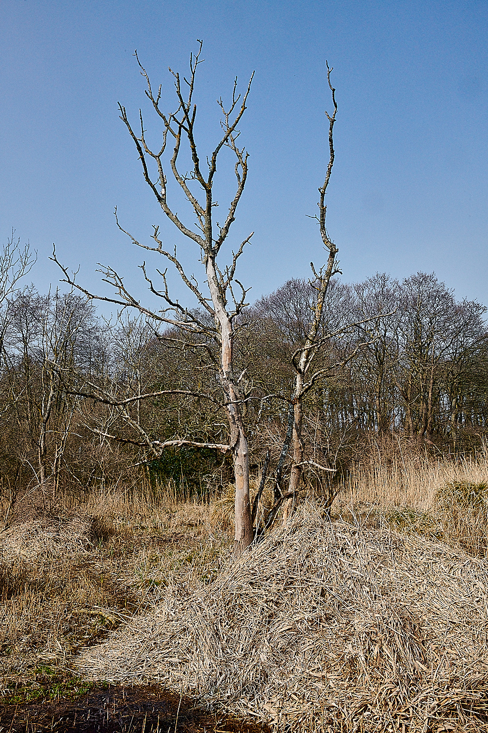
Fresh Reed Cutting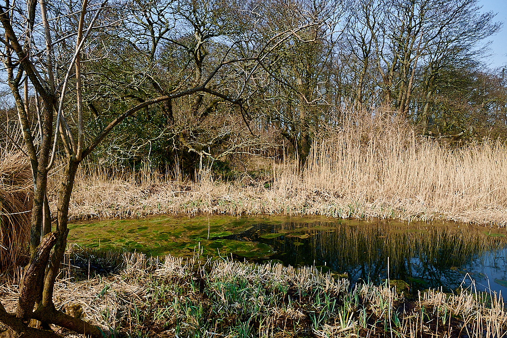
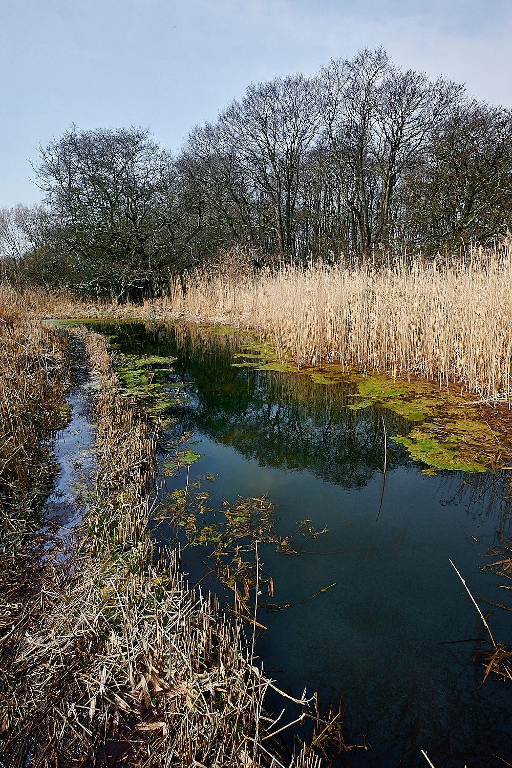
New ponds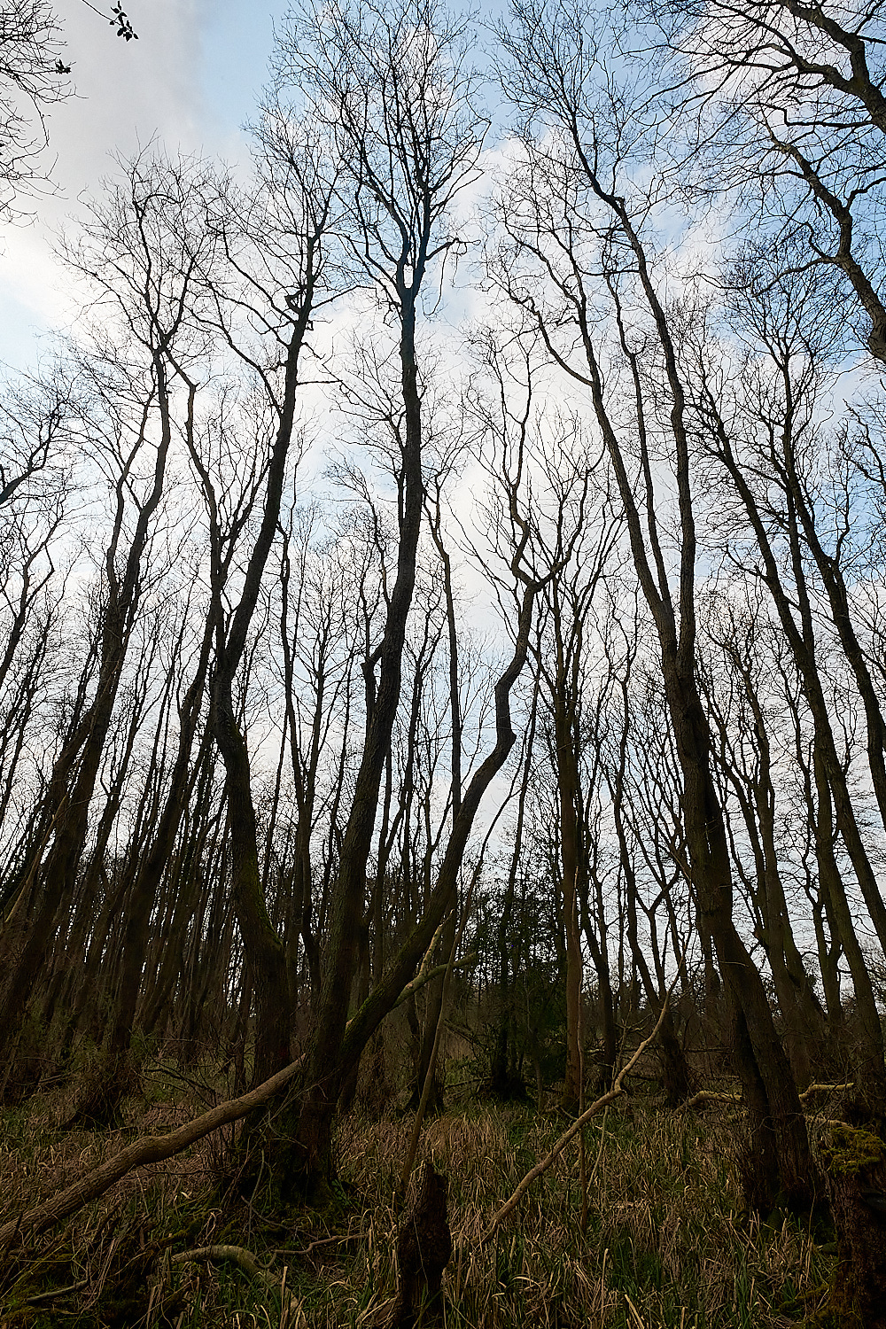
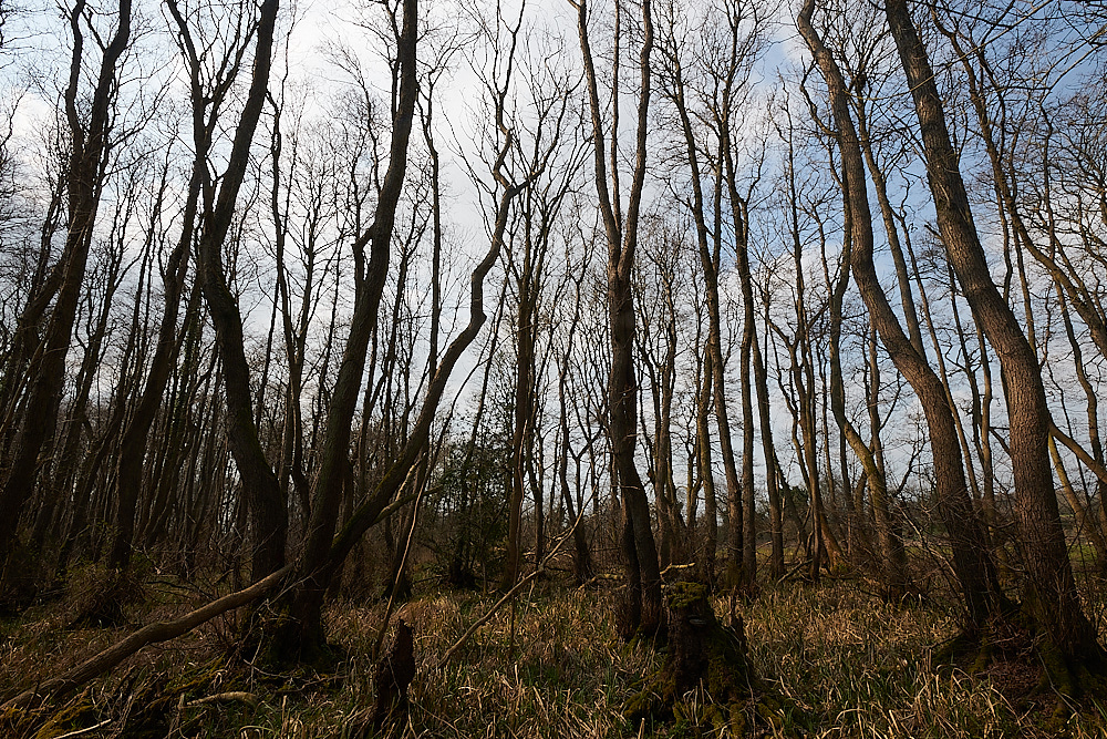
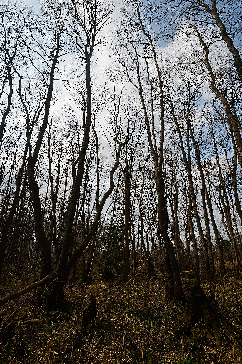
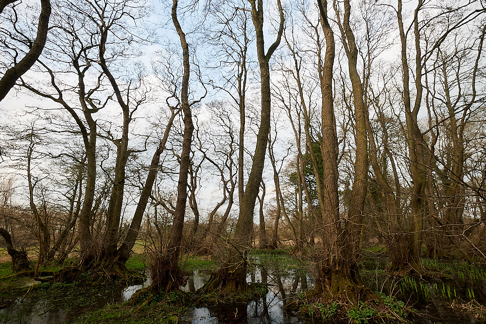
Alder (Alnus glutinosa) woodland. - About 40 years old.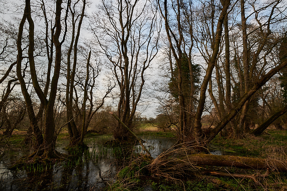
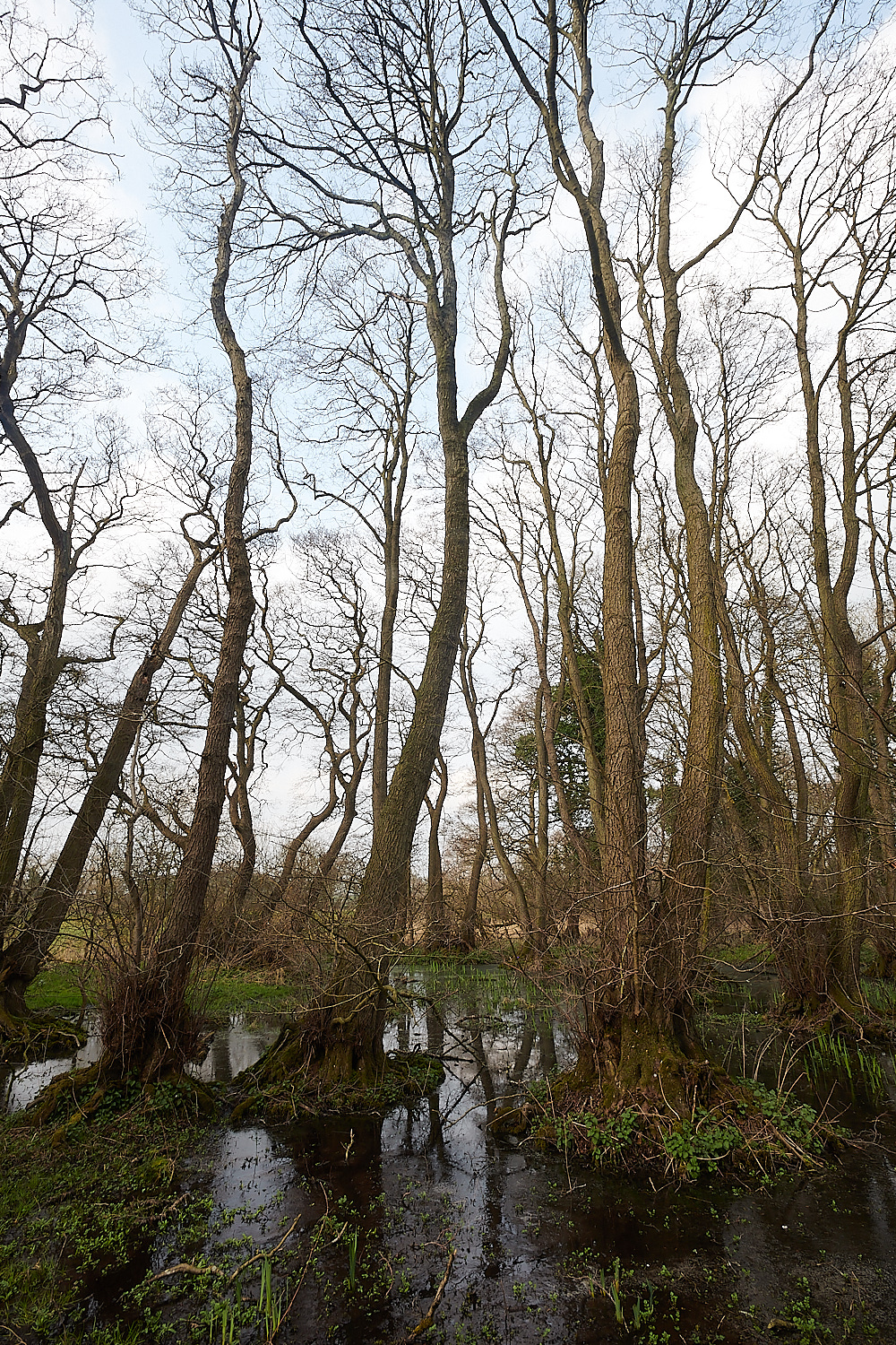
Older Alder 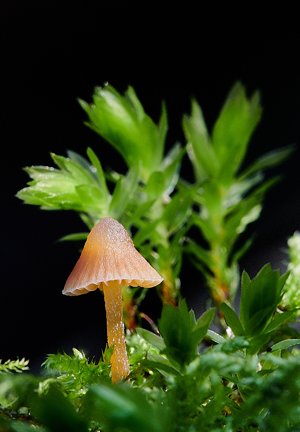
Galerina Sp with Swan's-neck Thyme-moss (Mnium hornum) in the background.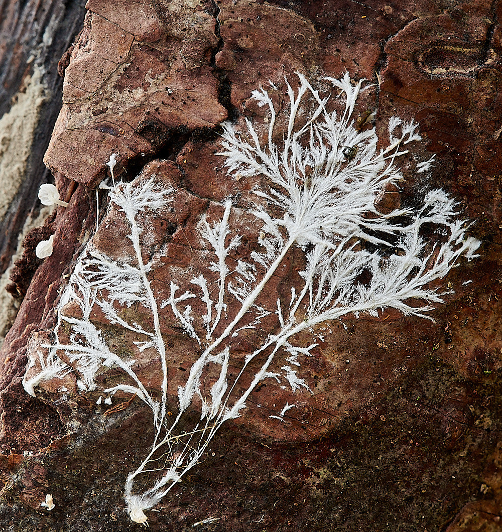
Mycelium pattern of Dewdrop Bonnet (Hemimycena tortuosa)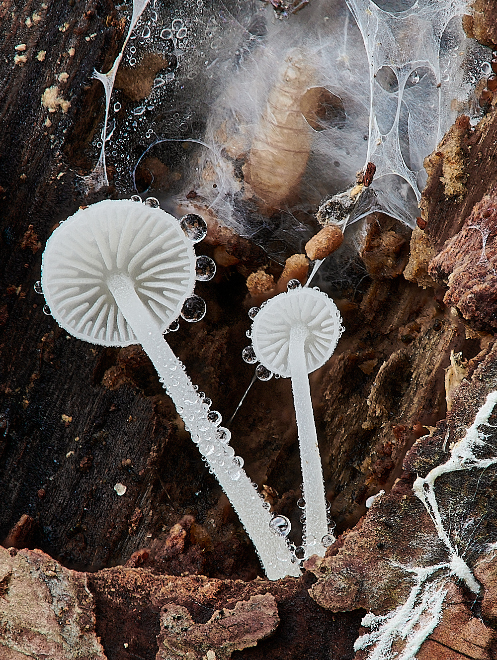
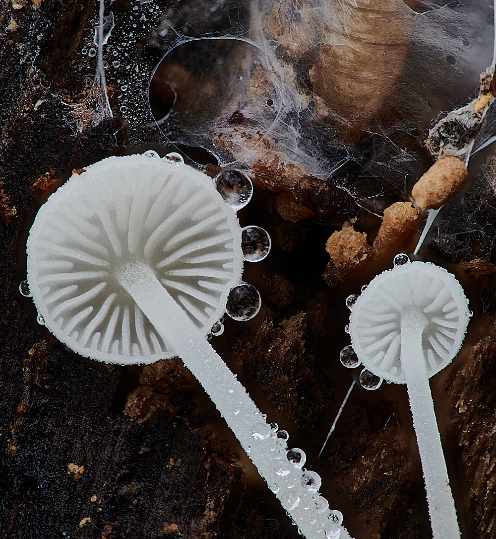
Dewdrop Bonnet
Mark the top one was taken with the 1.6 filter.
The bottom one was taken with the 1.6 Filter plus the Raynox 250 filter - giving 3.6 magnification.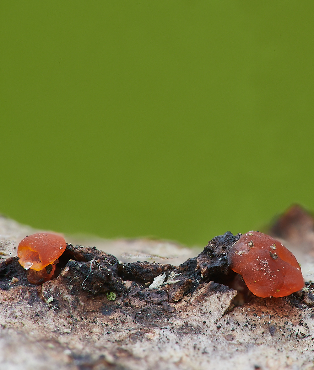
Fungus growing on a Woodwart Sp
To be identified
Mark - This one taken with the combined 1.6 & 2.5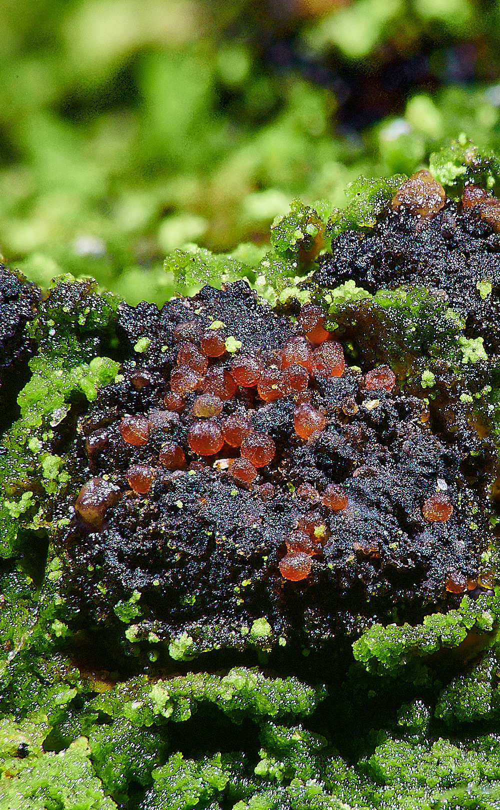
For comparison a Ruby Red microfungi also growing on a Woodwart Sp found at Upgate Common.
This one likely to be dialonectria epispheria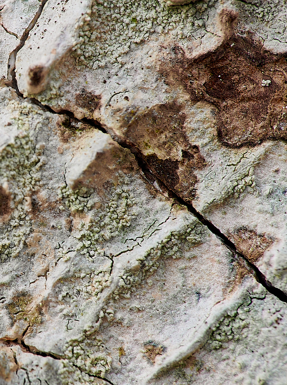
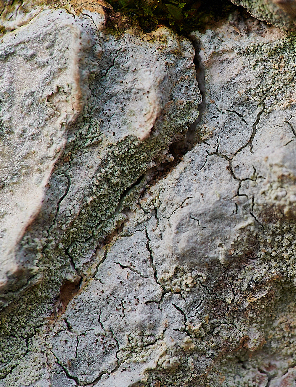
Lichen Sp growing on Oak.
Phlictis argenea
from the
British Lichen Society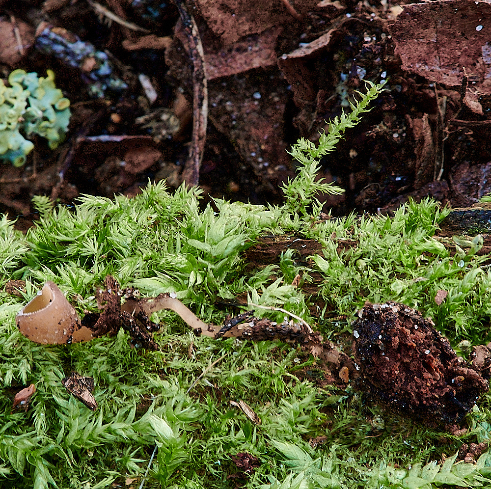
On the Hawthorn Berry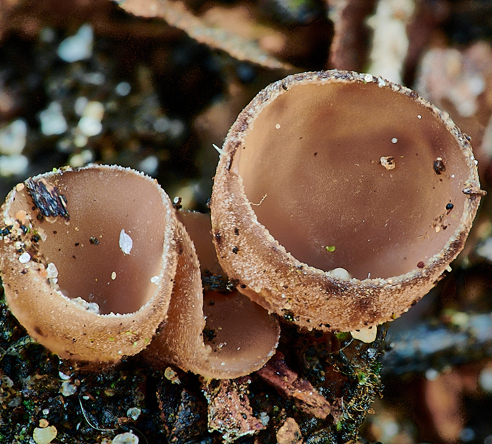
Haw Goblet (Monilinia johnsonii)
from
UK Fungi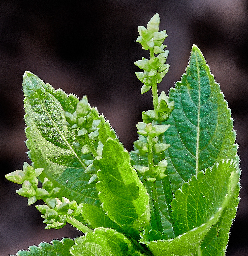
Dog's Mercury ( Mercurialis perennis)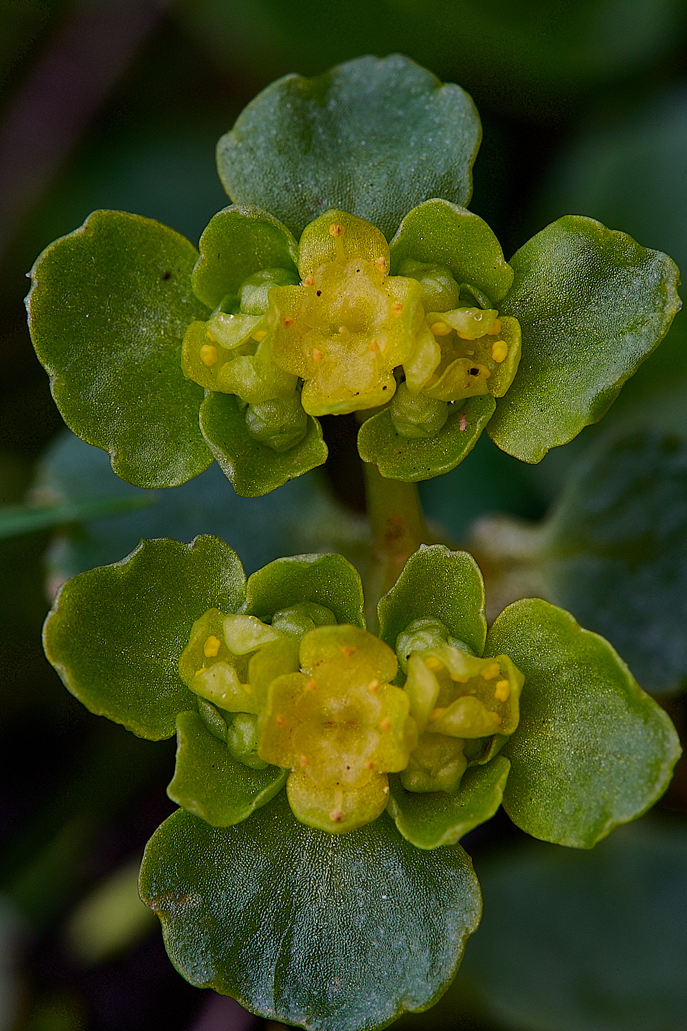
Opposite-leaved Golden Saxifrage (Chrysosplenium oppositifolium)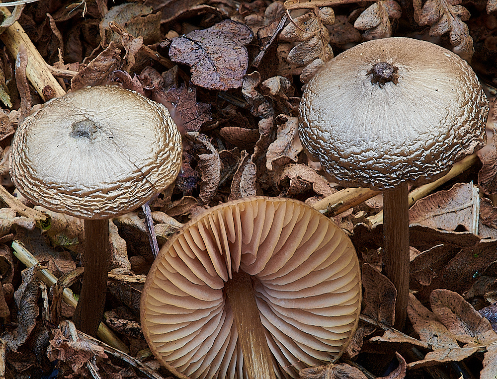
Pinkgill Sp Entoloma Sp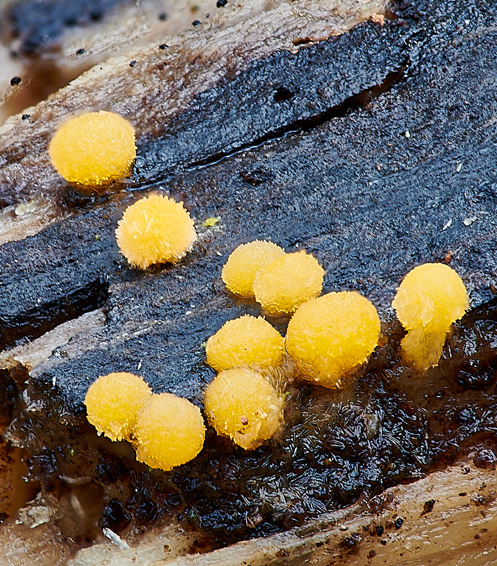
This one confused everyone.
To be determined.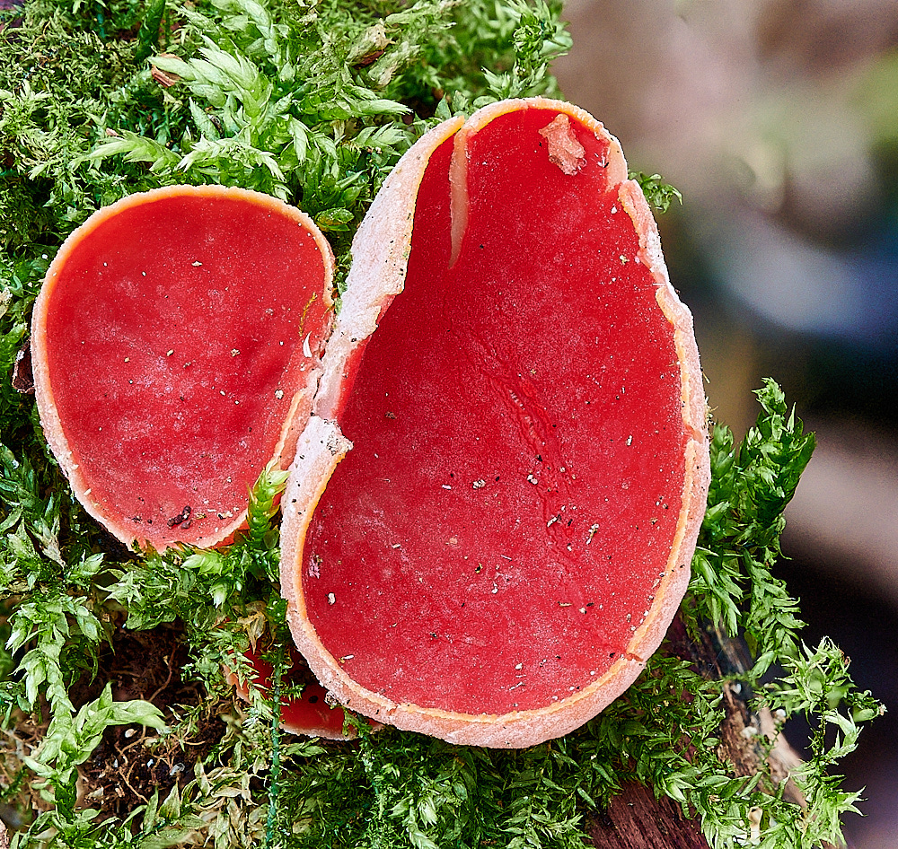
Elfcup Sp
from
Mark Jpy
Sarcoscypha austriaca, Scarlet Elfcup I found these on rotting fallen logs & thick branches (of Willow sp. I think) in moss.
Under the microscope the cup surface hairs were very colied (ruling out Ruby Elfcup, Sarcoscypha coccinea) & also the spores were too wide for Ruby Elf Cup).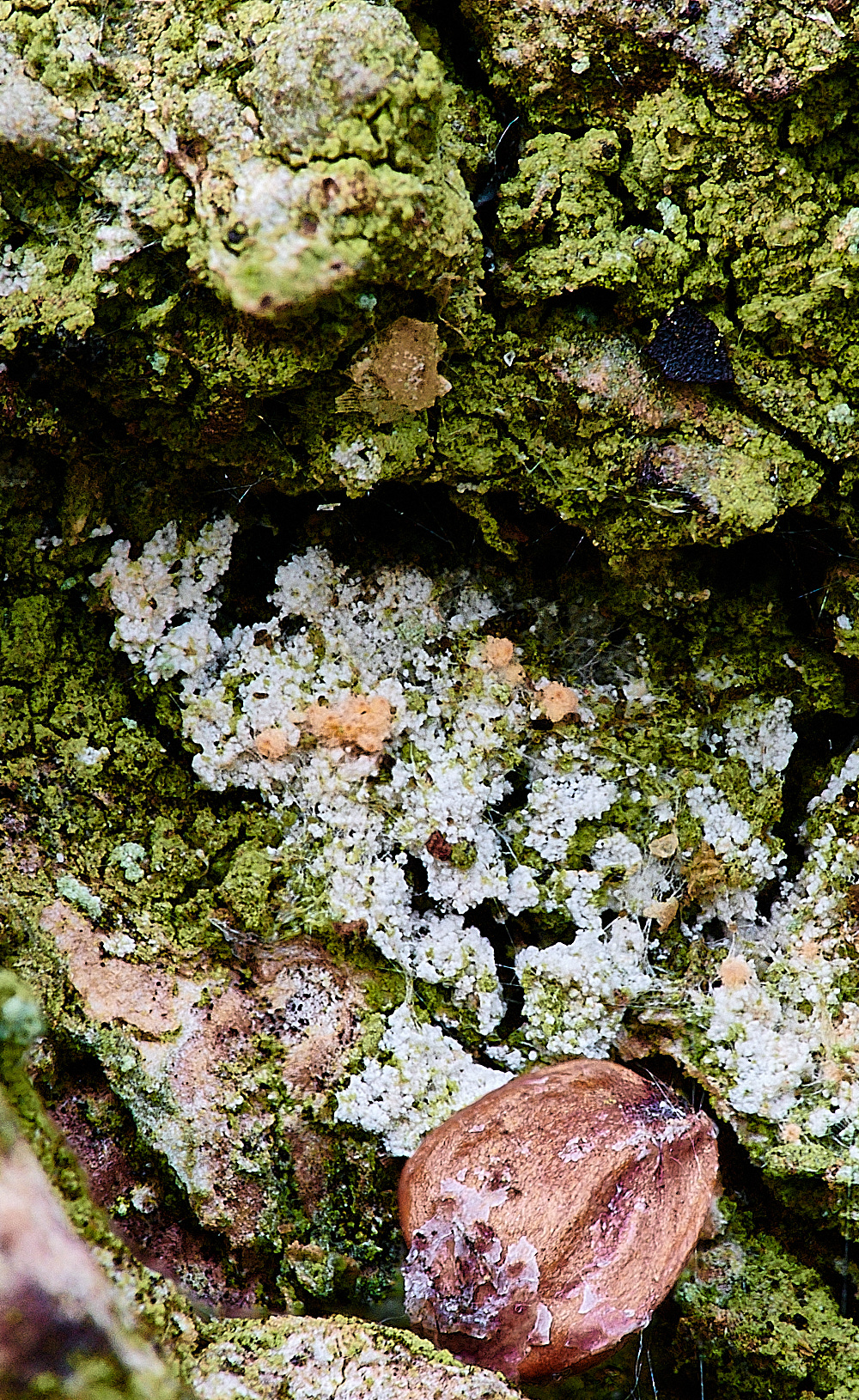
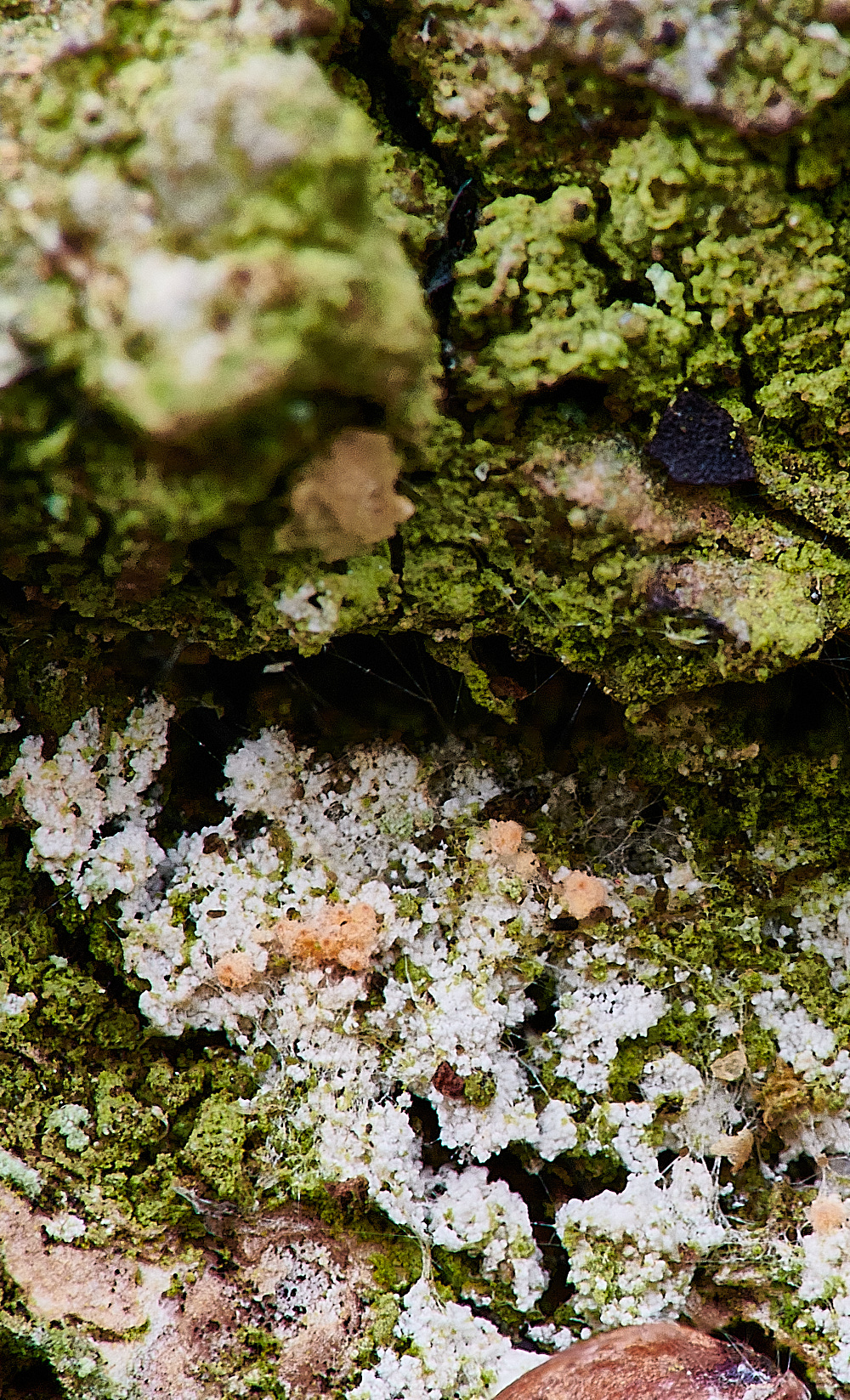
Fungus ona Lichen
Both to be determined.
Mark again the top one taken with the 1.6 and the lower one with the combined 1.6 & 2.5
Upgate Common
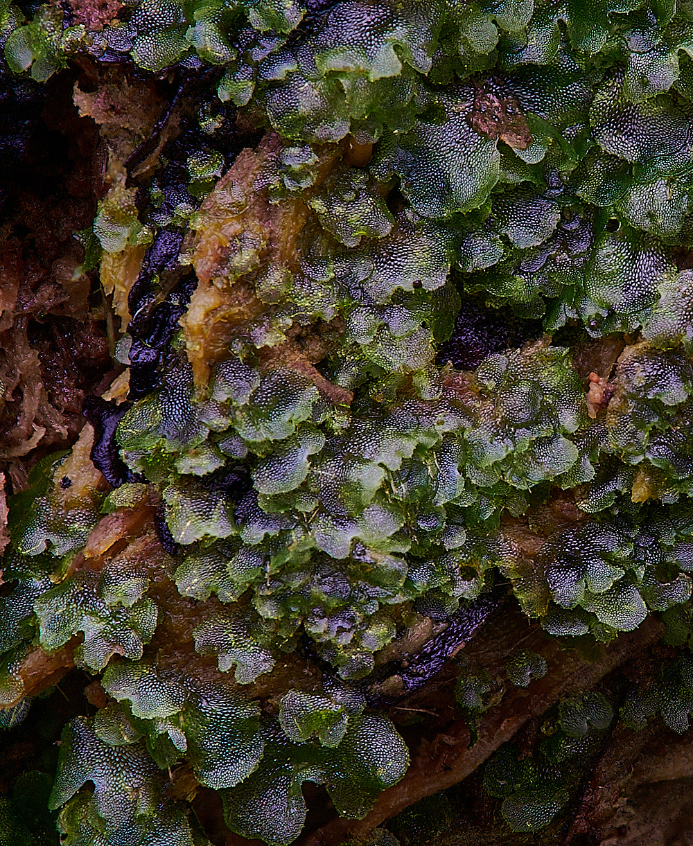
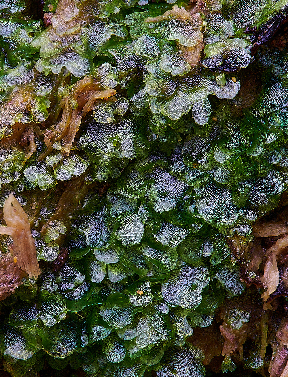
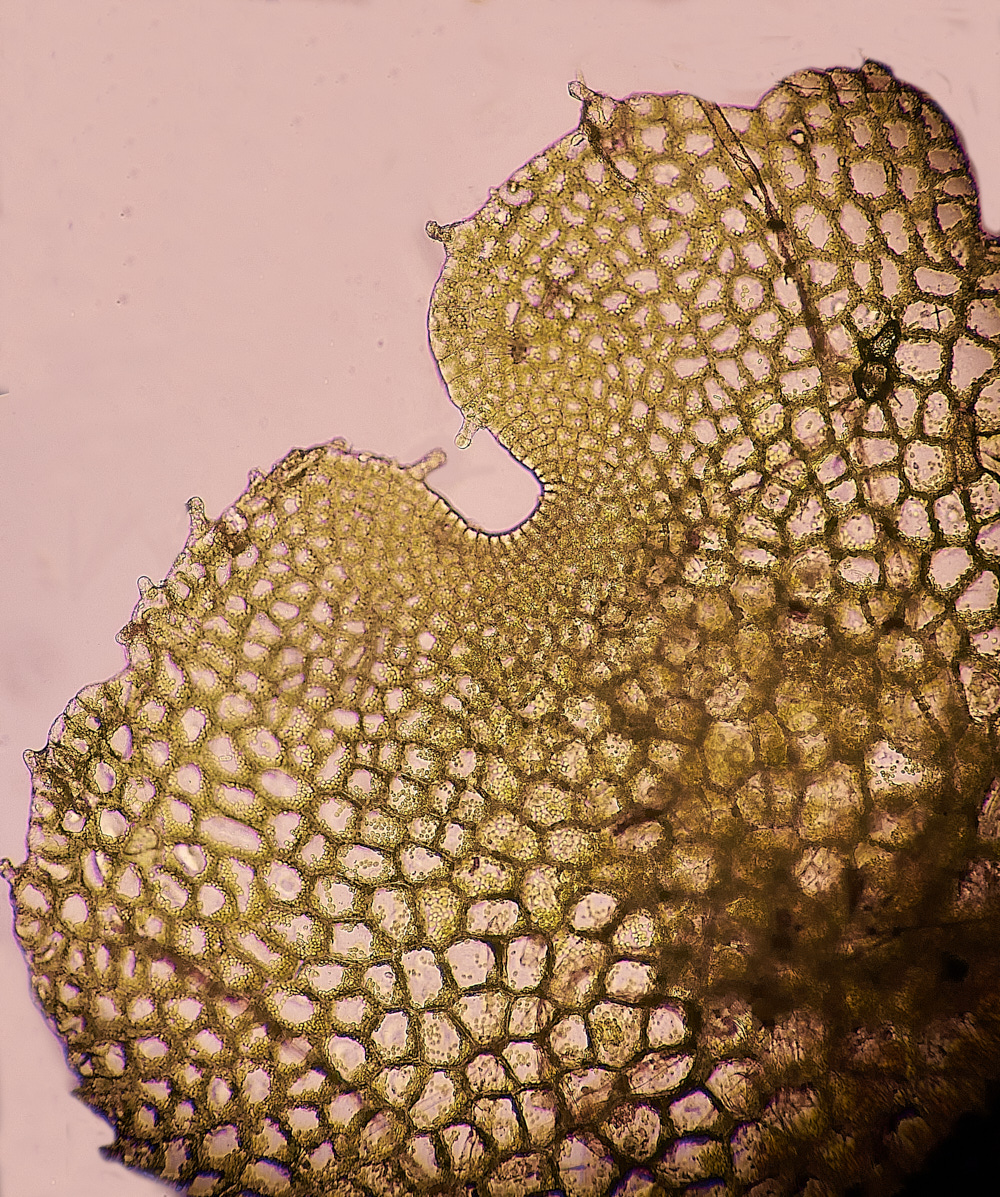
Common Pouchwort (Calypogeia fissa)?
That was certainly the first thought but it seems possible that this is something else.
Notice the little tags on the edges of some of the leaves and the notches are certainly large for this species. I also couldn't find a central midrib as it was very connected to the substrate.
I will pass this one on to Mary.
Actually
Fern prothalli - gametophyte stage of one of the Dryopteris species.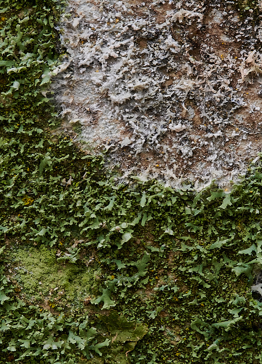
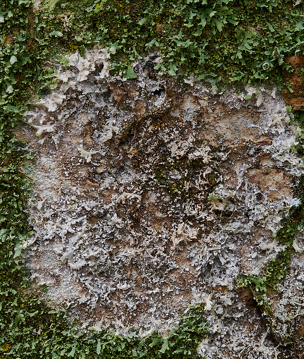
In this photo the centre of the white circle is dead and the Cobweb duster is moving out from the centre.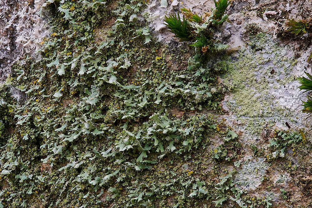
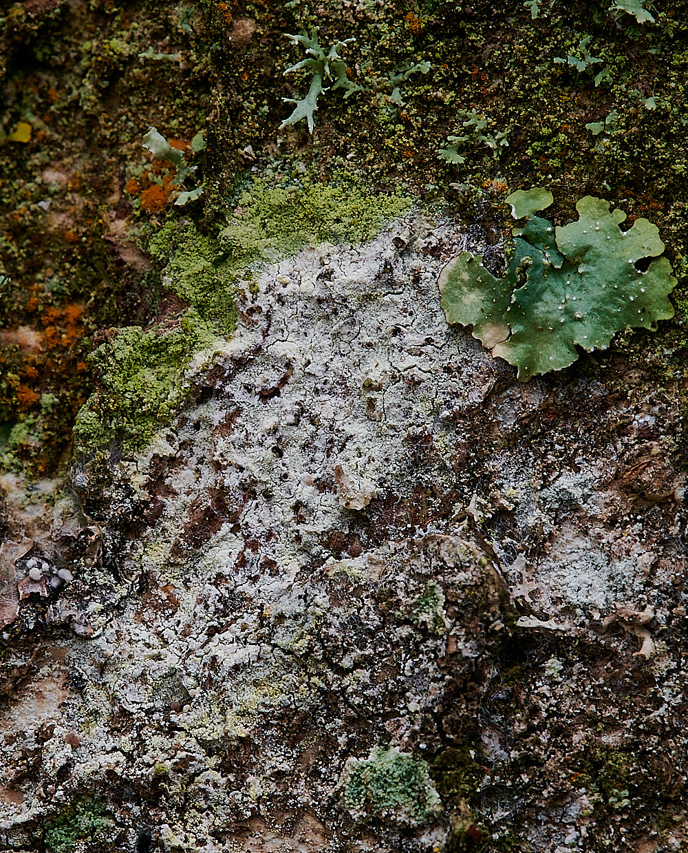
Cobweb Duster (Athelia arachnoidea) A corticoid fungus that forms thin white cobwebby basidiocarps that is a parasite on lichens.
From Mushrooms of Russia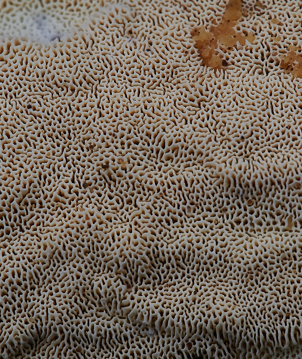
Common Mazegill (Datronia mollis)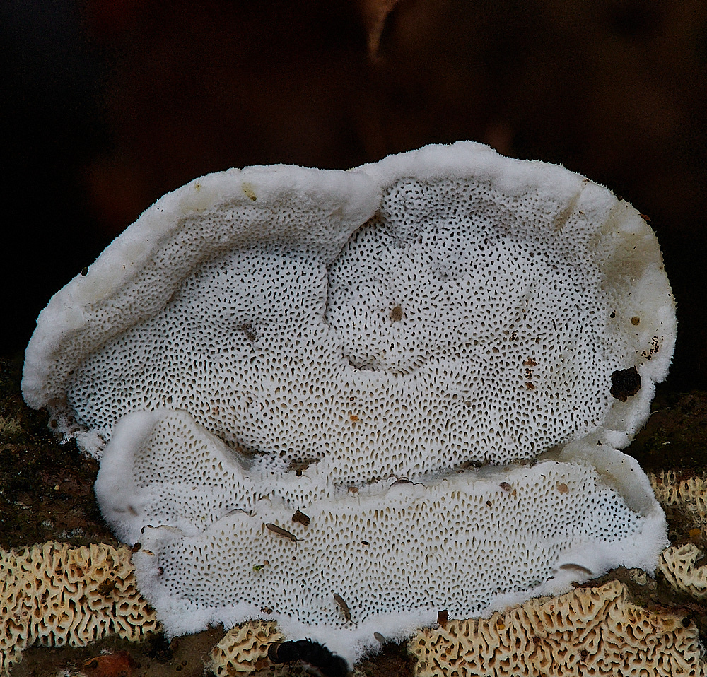
Fungus Sp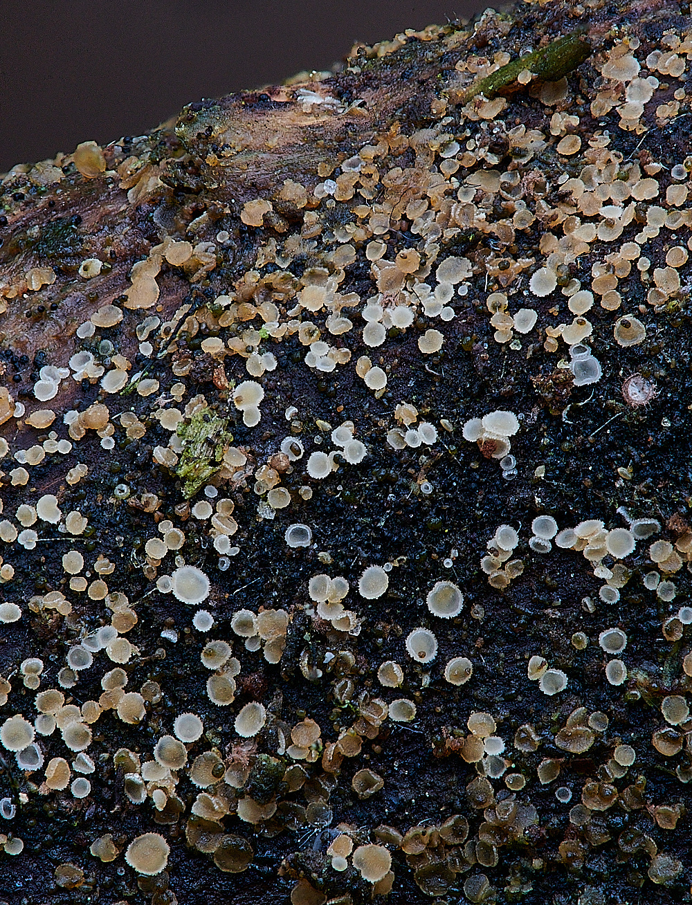
Disco community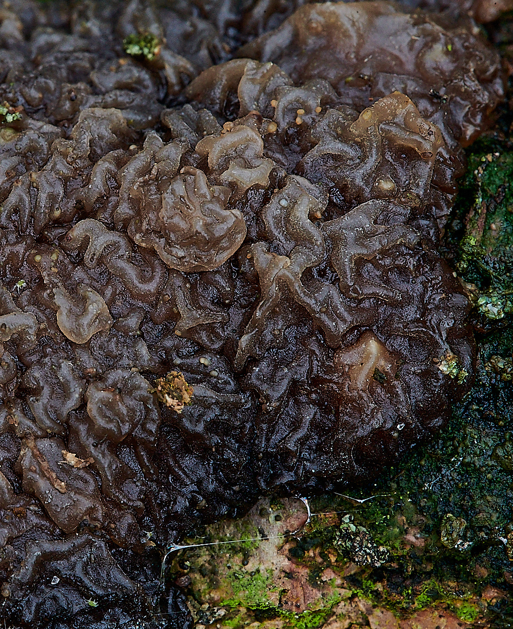
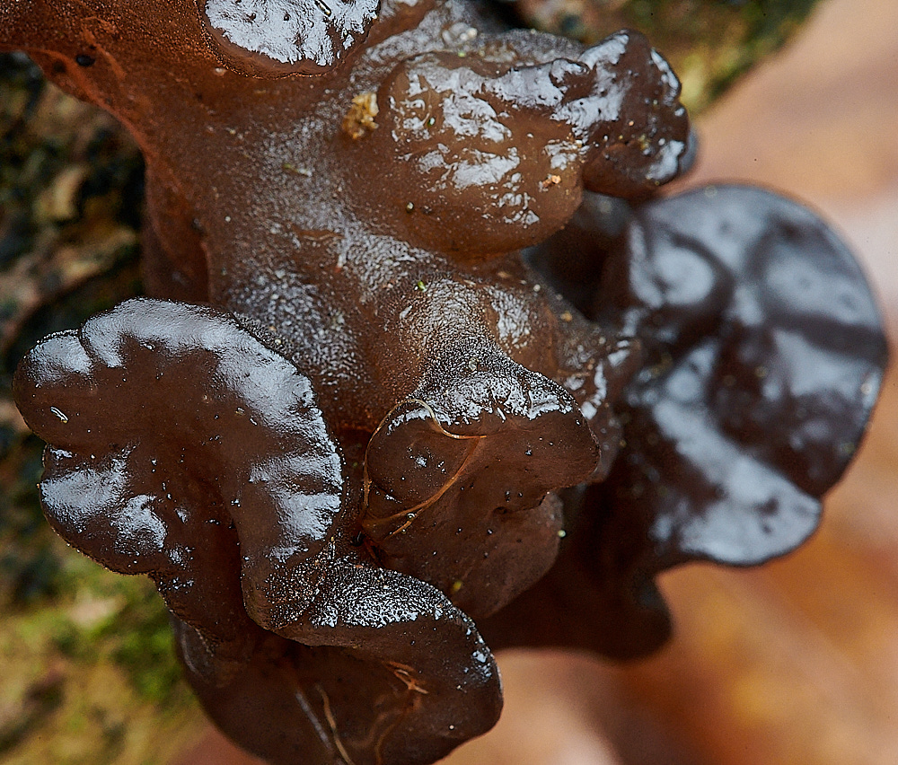
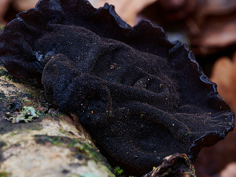
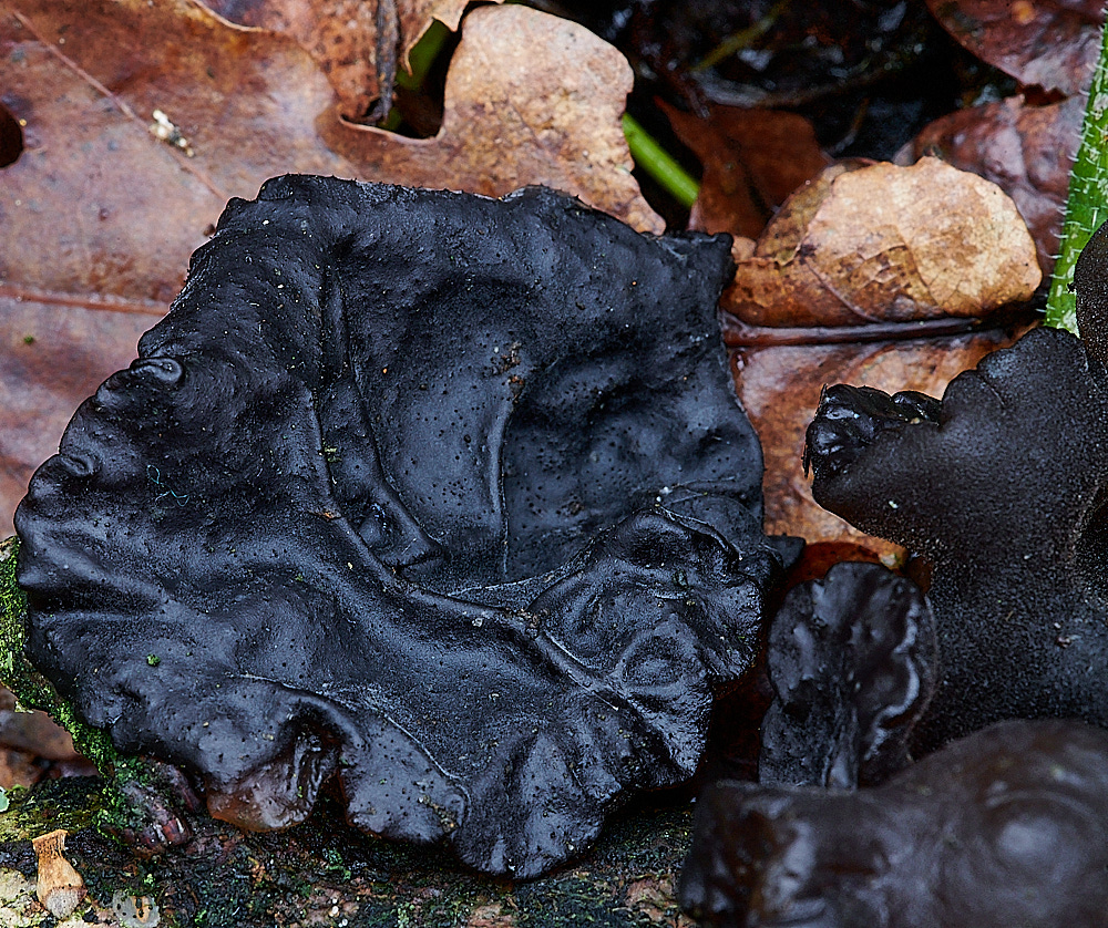
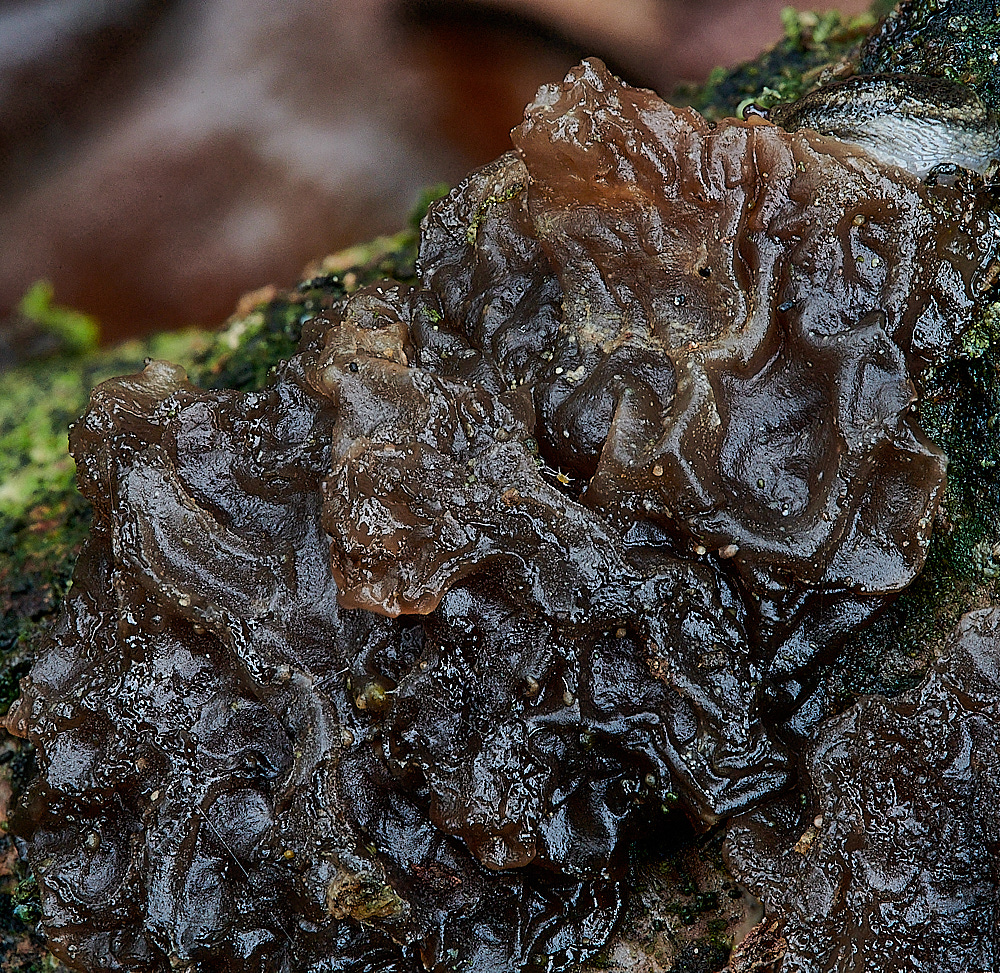
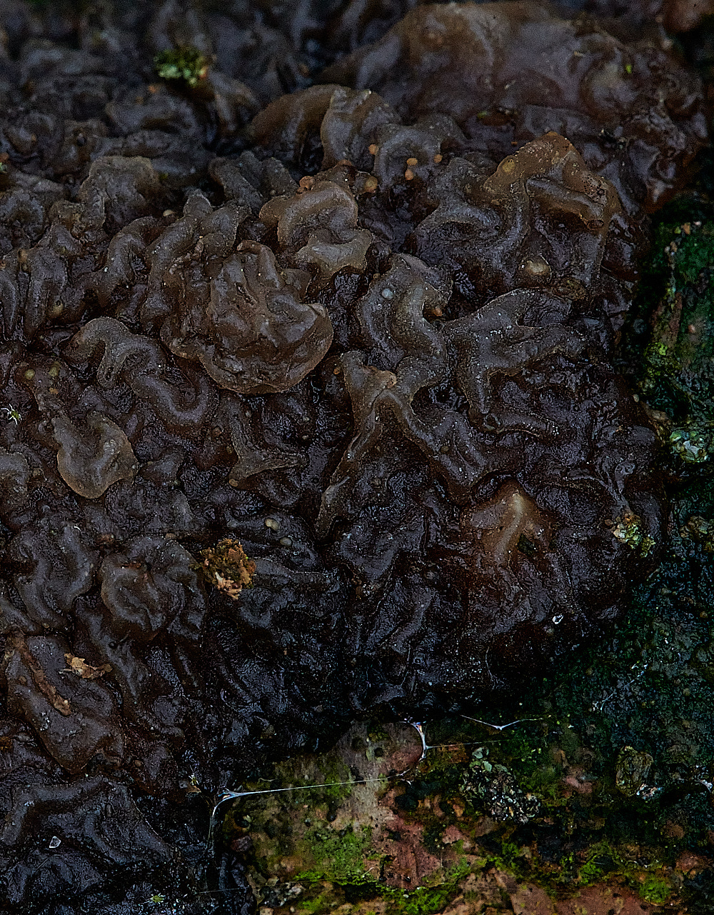
Witches' Butter (Exidua Sp)
Possibly two different forms. The one a very dangly, gangly form, the other very closely adpressed to the branch. Both very close to each other.
One a very mature looking specimen with an almost velvet appearance on one side.
A head scratching mystery for further study.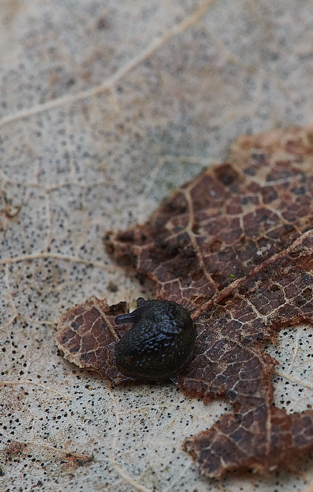
Hedgehog Slug (Arion intermedius)
Lichen Sp?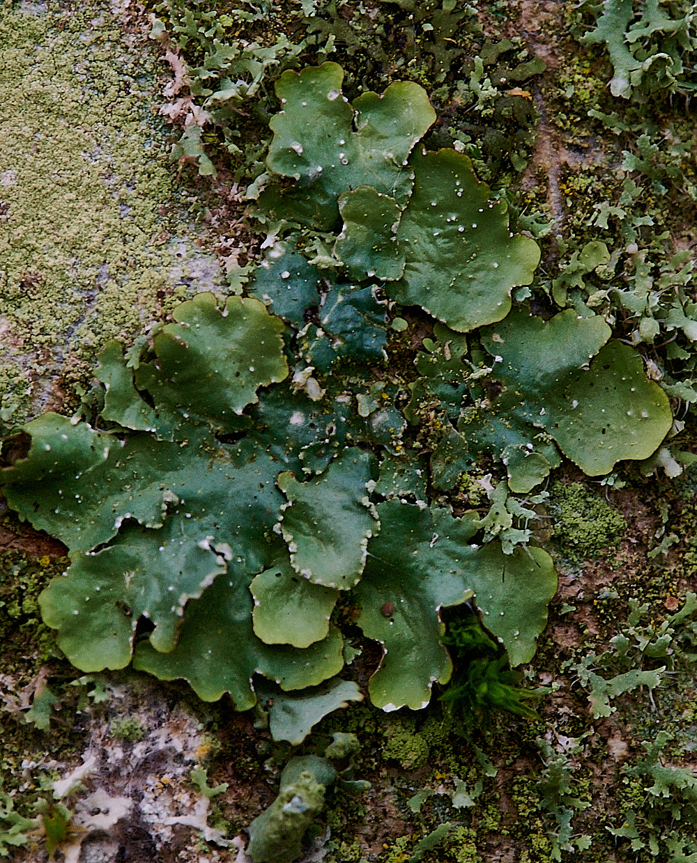
Lichen Sp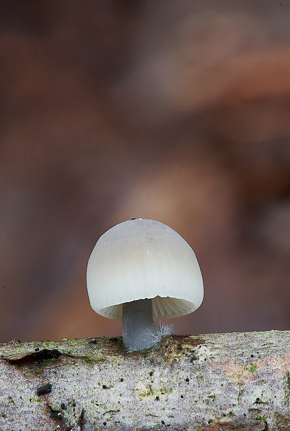
Angel's Bonnet (Mycena arcangelica)
The cheilocystidia were of the pear form with lots of bristles - Hedgehogs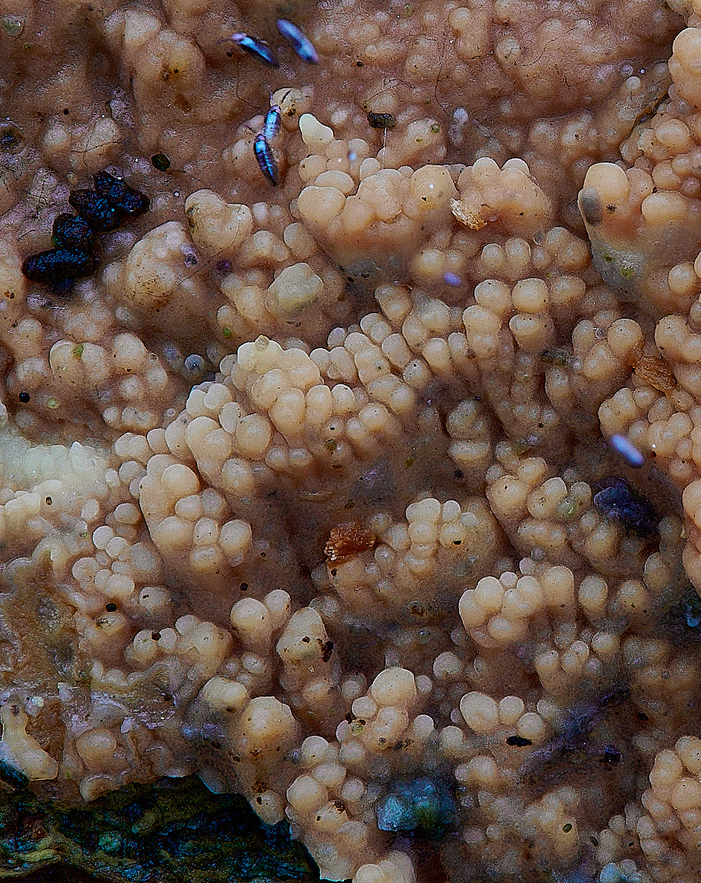
Possibly a strange form of Wrinkled Crust (Phlebia radiata)?
from Anne
But I was stuck with the small spored toothed fungus that we thought looked a bit like a Phlebia on site and have just discovered it might be Hyphodontia quercina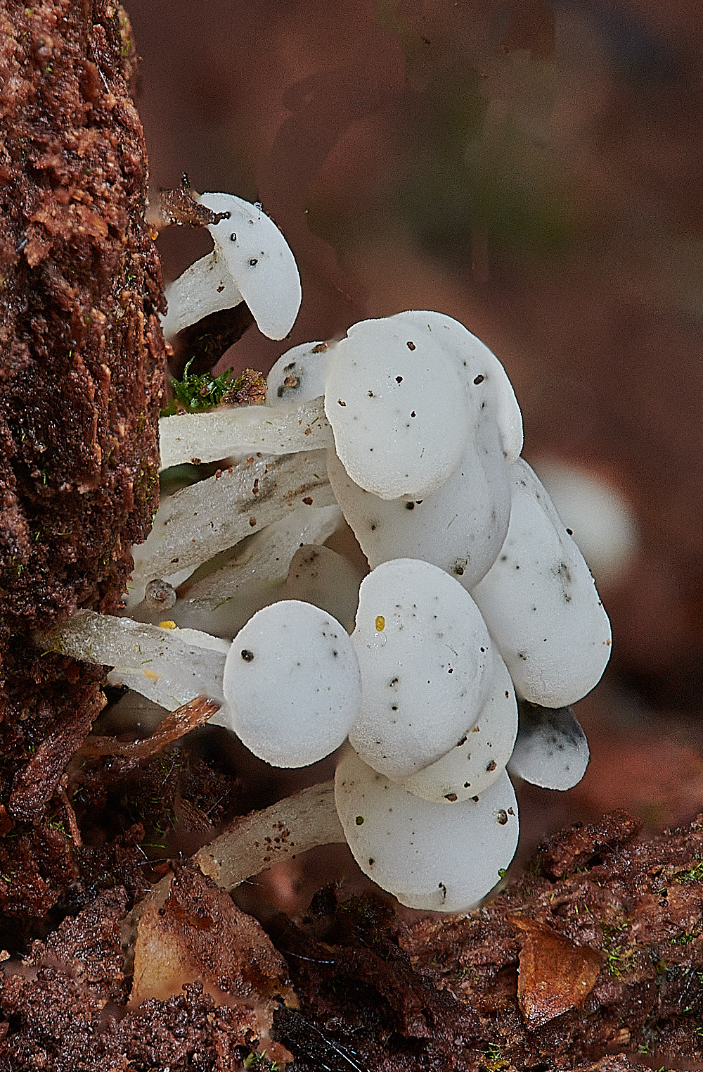
A white pin type fungus on Alder ( Alnus glutinosa)
from Steve
The little white things on the dead and very decayed stump turned out to be Cudoniella acicularis - Oak Pin. Ascus, spores (many 1 septate) and paraphyses measured and all fitting nicely within the required parameters. (Fungi of Switzerland, Peter Thompson Ascomycetes and Fungi of Temperate Europe)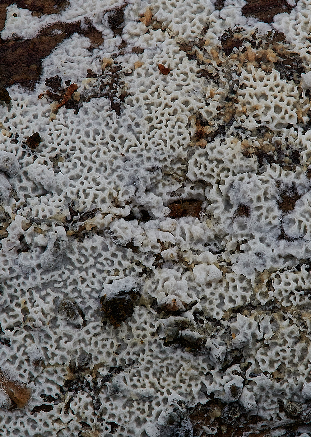
Poroid Fungus?
from Anne
The small honey comb-like resupinate that I was hoping would be a Trechispora was Ceriporia reticulata.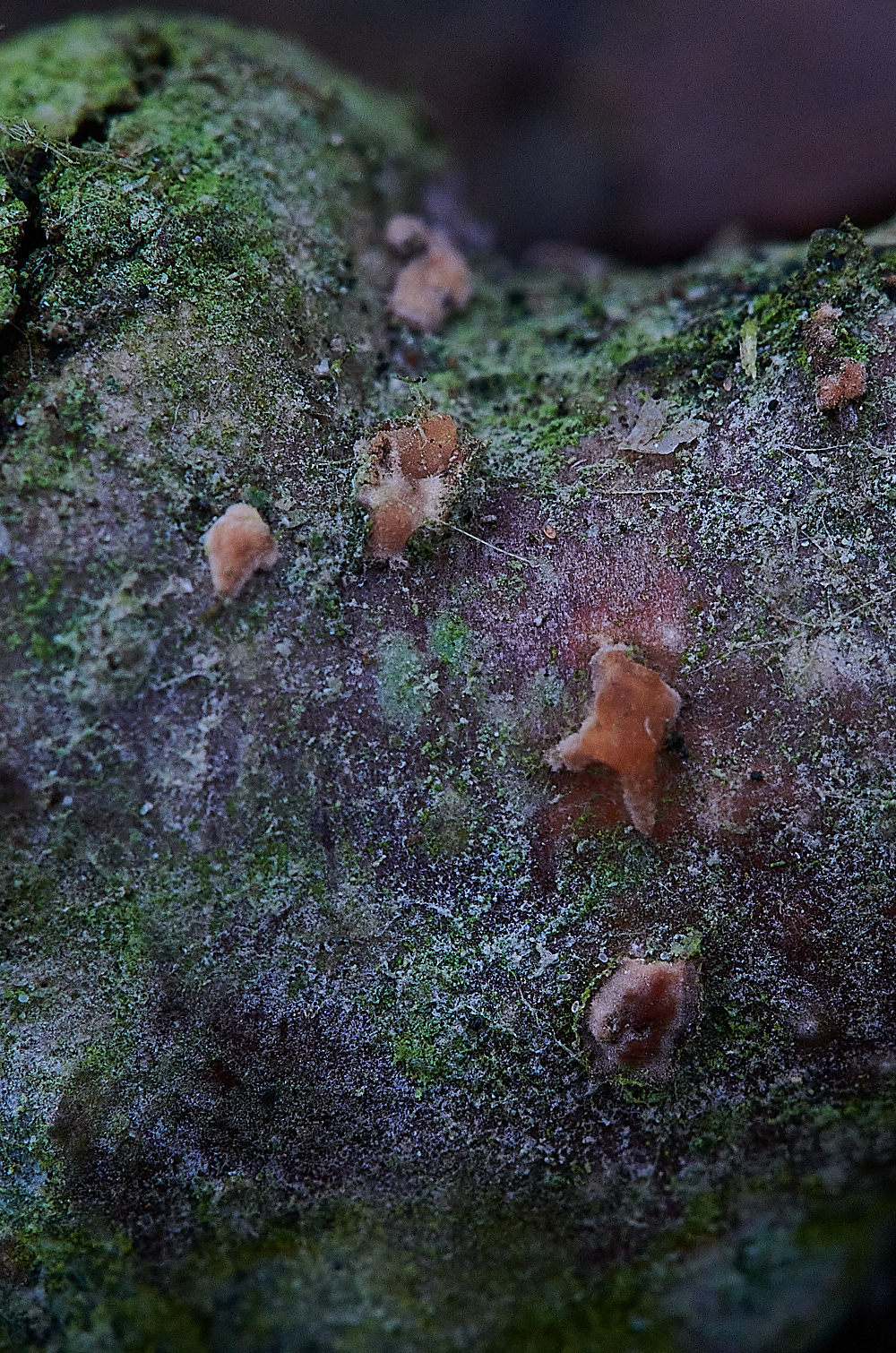
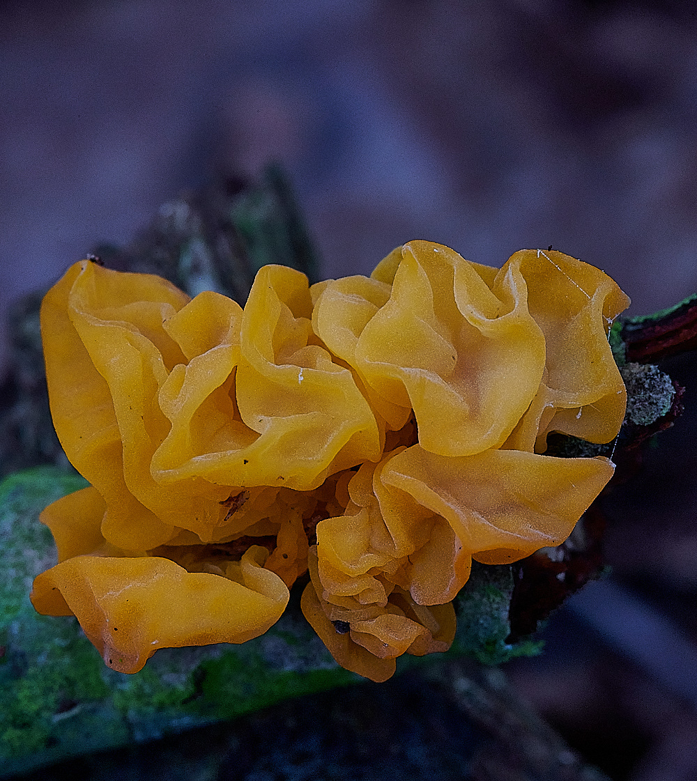
Yellow Brain (Tremella mesenterica)
From First Nature
Yellow Brain fungus grows on dead wood that has been attacked by wood-rotting fungi of the Peniophora genus. One of the most common Peniophora crust fungi in Britain and Ireland is Peniophora incanata, commonly known as Rosy Crust fungus. Very little or none of the Peniophora may be visible; this is because Tremella mesenterica feeds on the mycelium of the Peniophora fungus, and that can be deep inside the timber rather than on its surface. The fruiting body of the crust fungus does not even have to be present, therefore, and so it may look as though Yellow Brain is feeding directly on the host wood.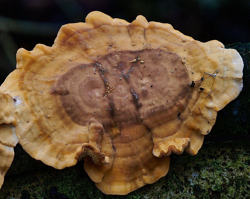
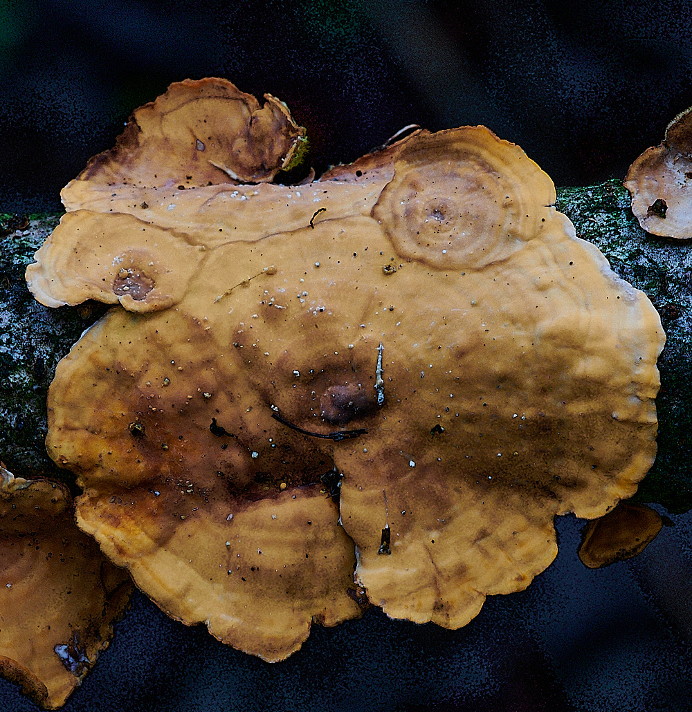
Yellowing Curtain Crust (Stereum subtomentosum)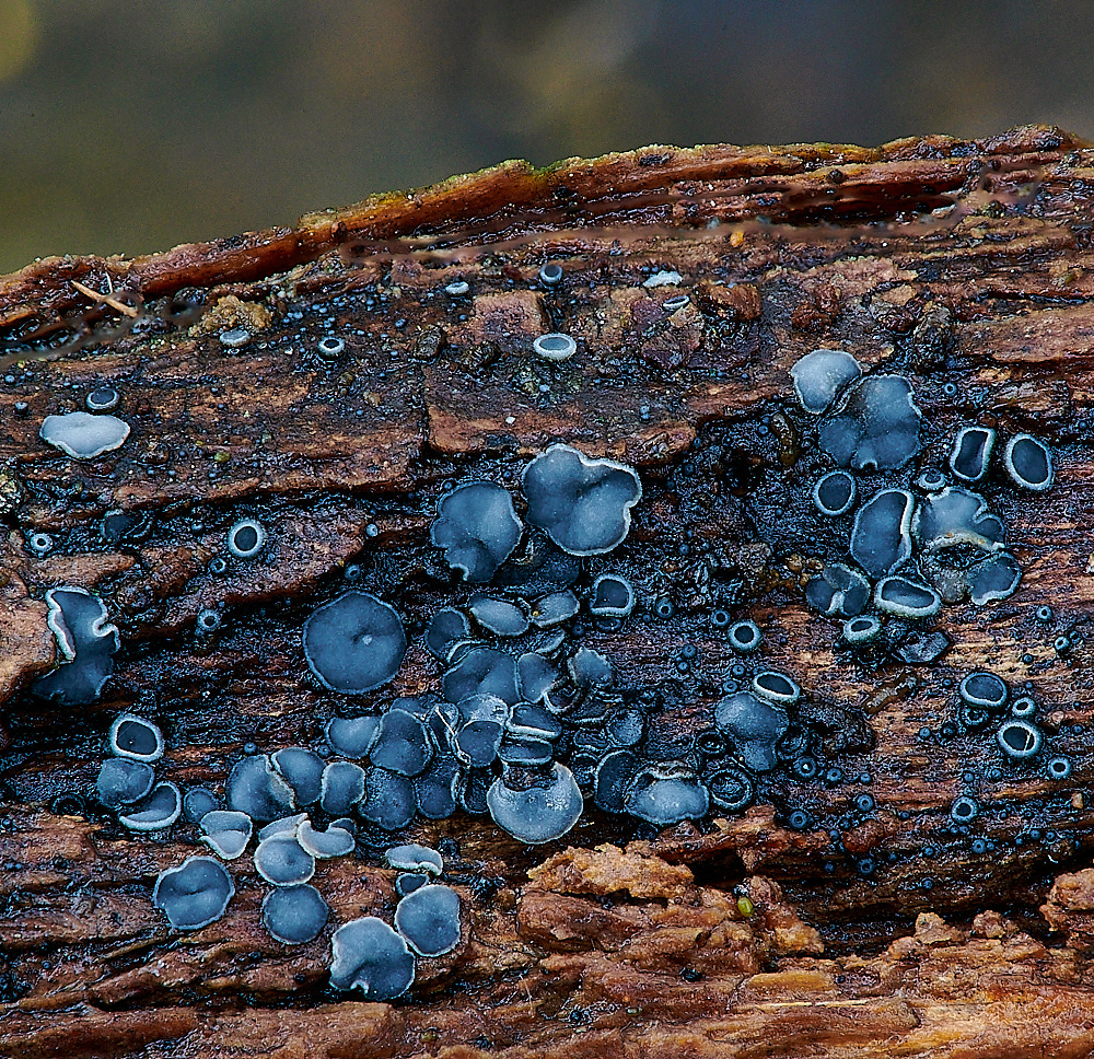
Common Grey Disco (Mollisia cinerea)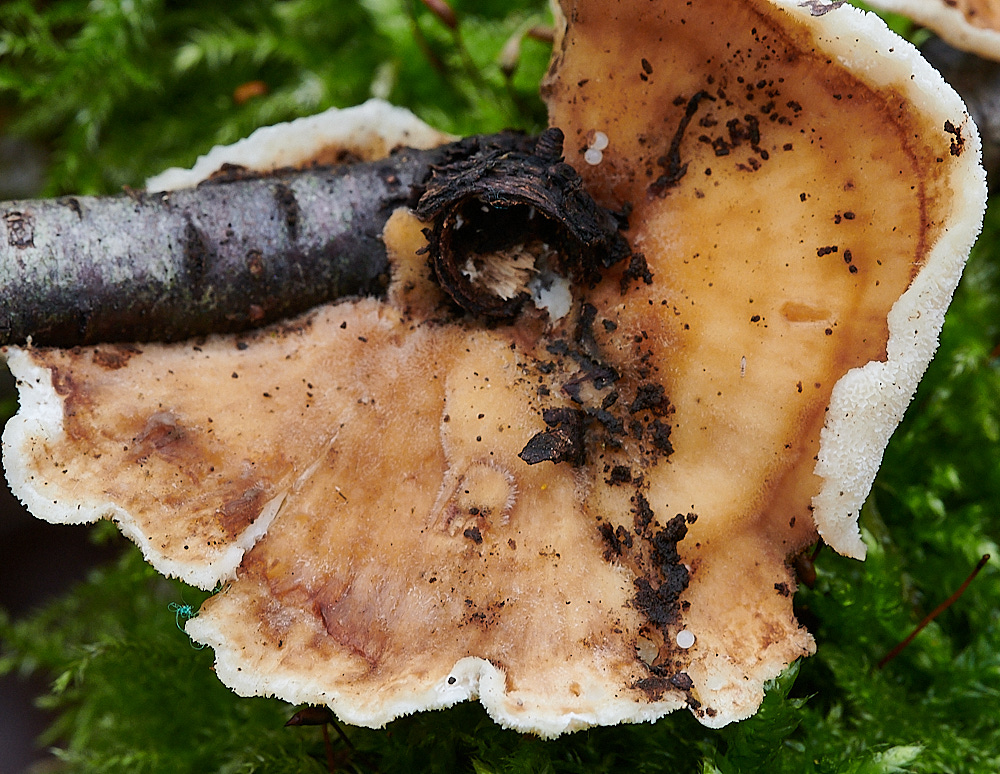
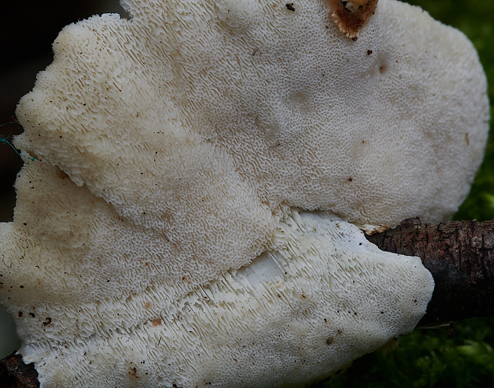
Turkey Tail
Possibly Ochre Bracket (Trametes ochraceae)
became
Trametes pubescens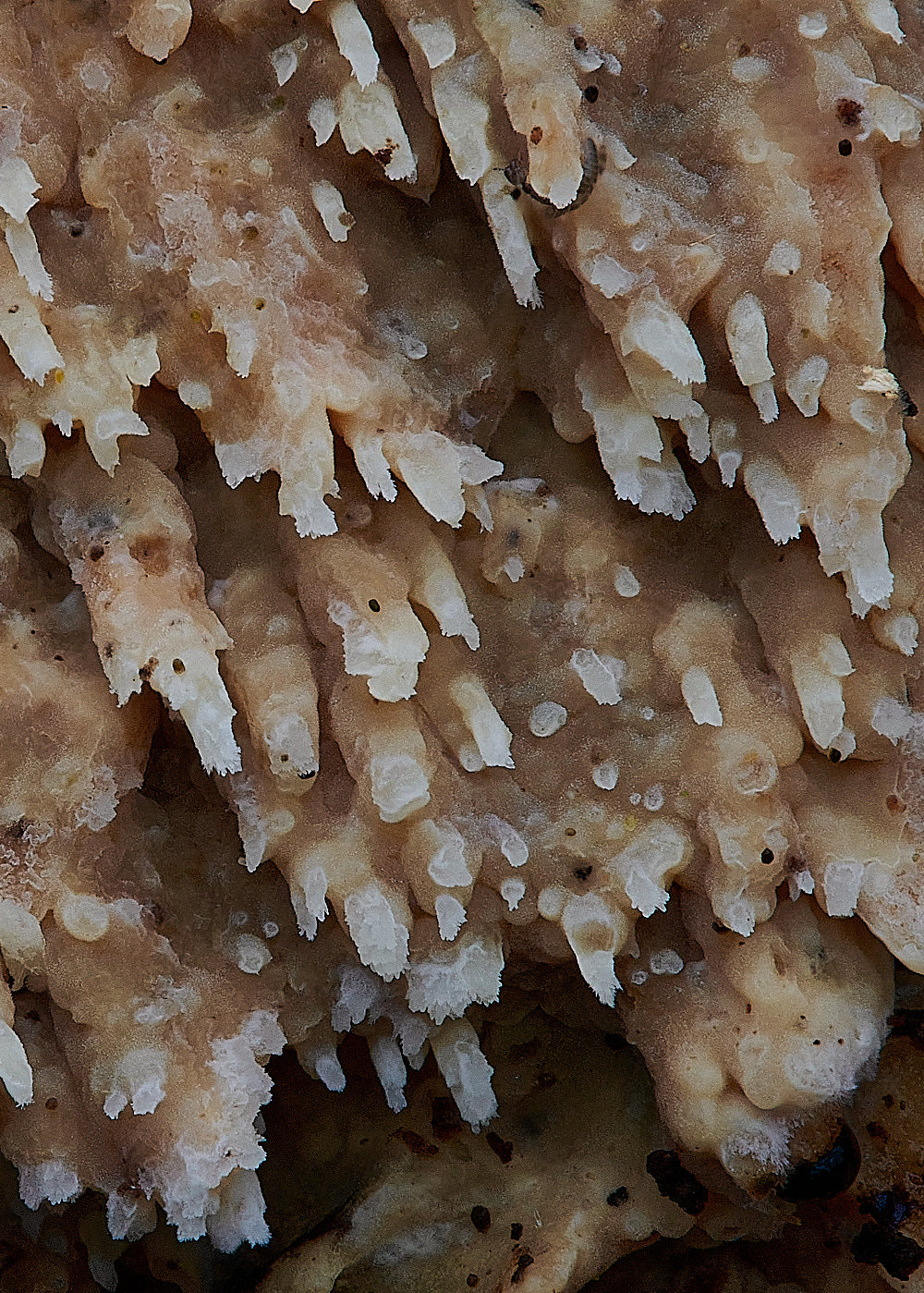
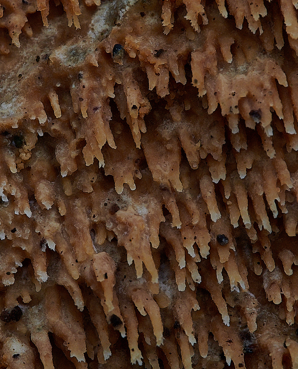
Could be one of two
Oak Tooth Crust (Radulomyces molaris) or Toothed Crust Fungus (Basidio radulum radula)
In this case it turned out to be Oak Tooth Crust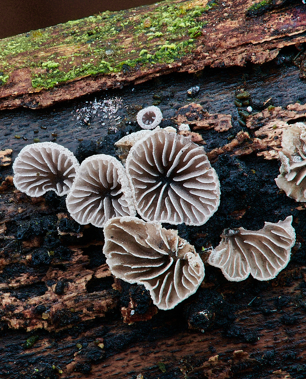
Smoled Oysterling (Resupinatus applicatus)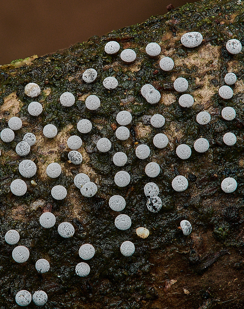
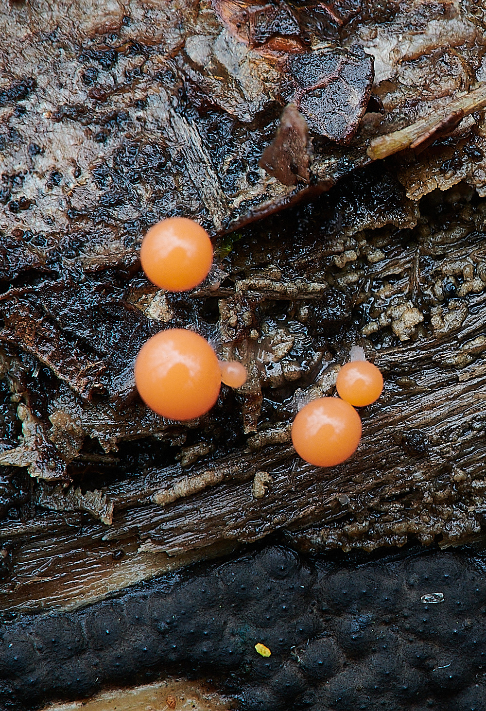
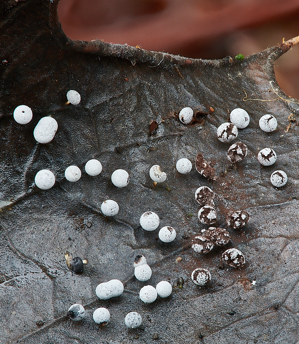
Slime Mold Species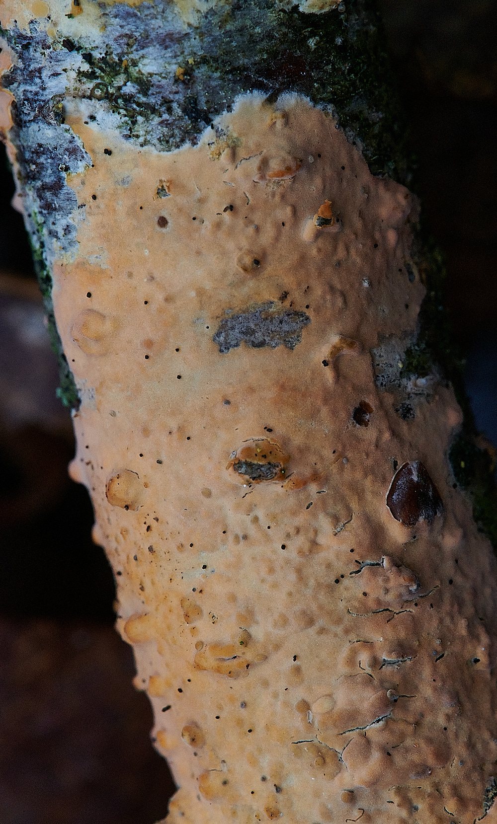
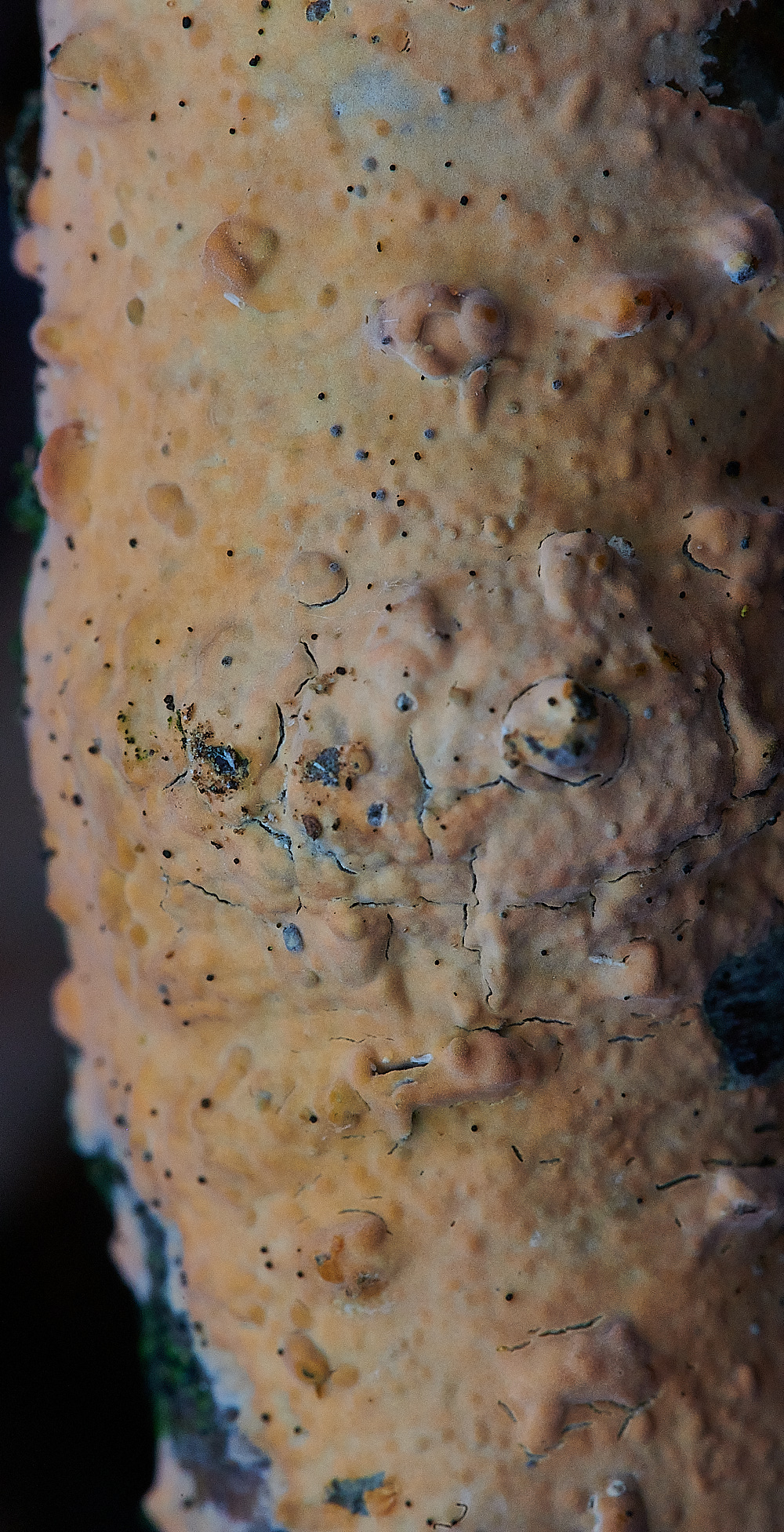
Rosy Crust (Peniophora incarnata)
But no
From Tony
I thought that the orange crust (SJ took some too I think) might be Rosy Crust Peniophora incarnata but the spores were too small (5x3) & cystidia didn’t look right. In fact I couldn’t find any encrusted cystidia at all so perhaps not a Peniophora after all.
But an initial injunction from Steve
'On the fungal front a piece of advice, if anyone offers you a twig covered in a lovely orange pink peniophora like resupinate, do not accept it, run, do not look back and don't stop running.
Turned into a four day marathon
Having been working on the orange crusty thing on a stick for the past four days I am at last satisfied that we can add Peniophora incarnata to the list.
Spores fitting within the range given in Fungi of Temperate Europe.
Clamps found fairly easily but an absolute nightmare to find any Lamprocystidia or Gloeocystidia, 19 slides of samples and only one measly pointy thing found that might, with a hefty dose of imagination, possibly be a Gloeocystidia.
Decided to try something completely different today by taking my sample from the very edge of the fungal growth, and there they were Lamprocystidia aplenty, whilst more difficult to find there were Gloeocystidia as well both looking just like they do in the books.
I will confess to being on a bit of a high in having finally cracked it, whilst at the same time being in awe of the fact that Anne does these resupinatey things on a regular basis, massive respect to you.
and then
Determination is clearly as valuable a tool as the microscope and books :-)
Were I to attempt another Peniophora, and I guess I might be tempted if only to see if they get easier, and were you to feel inclined to attack one, I made a possible interesting discovery. As you probably picked up from the emails both I and Tony M. tried to get either Lamprocystidia or Gloeocystidia and there were none to be found apart from one measly possible specimen that I thought might have been. As I have mentioned out of sheer desperation I tried a piece from the very outer edge of the fruiting body and there were loads, I passed this finding on to Tony who still had a stick and he tried the same and immediately found them.
This rather begs the question of is this 'cystidia near the outer edges of the growth' typical of Peniophora, that will have to be a question for Anne next time I see her.
Anyway thought you might like to see a couple of photos of the results. The Lamprocystidia in the first photo is very clear protruding more or less in the middle of the photo.
In the second photo there are a lovely line of Lamprocystidia along the top edge, this photo shows just how easy they were to find once you got the right piece of material, there are also a couple of nice Gloeocystidia at the bottom mid right as well.
The third photo is the same view as the second but at a higher magnification and giving much closer views of both types of cystidia. 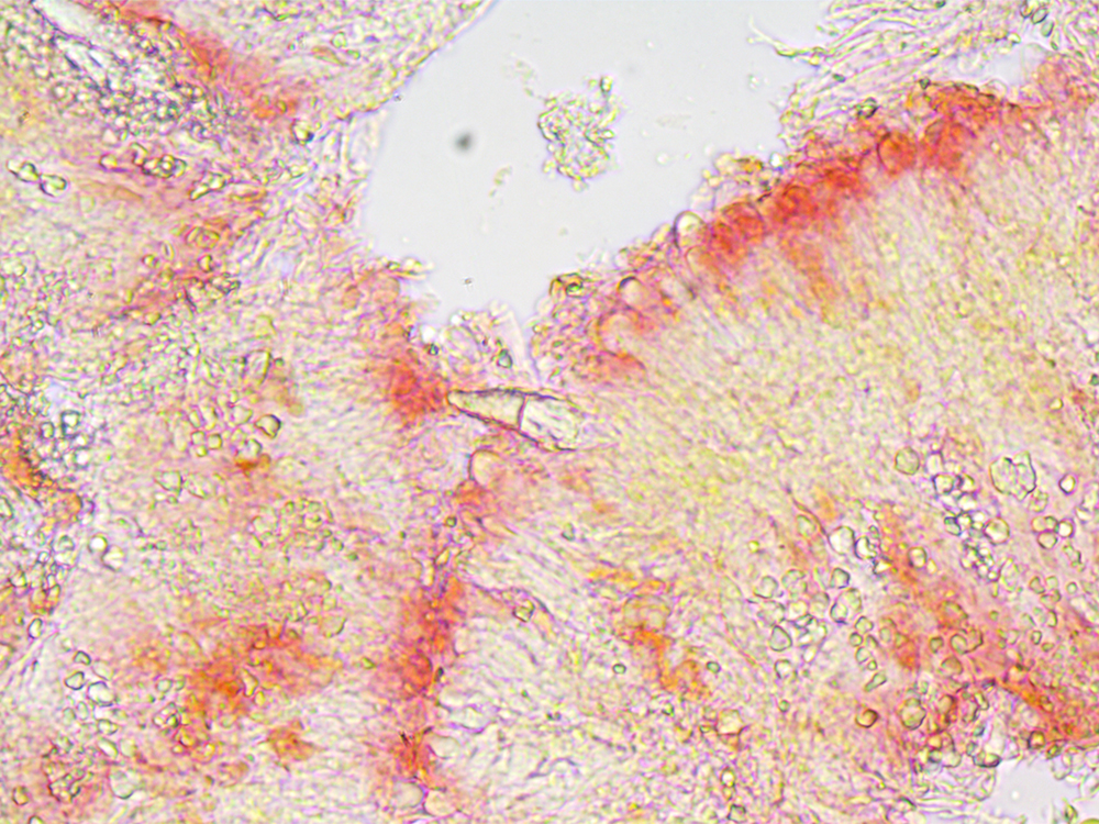
Lamprocystidia visible in the centre of the image.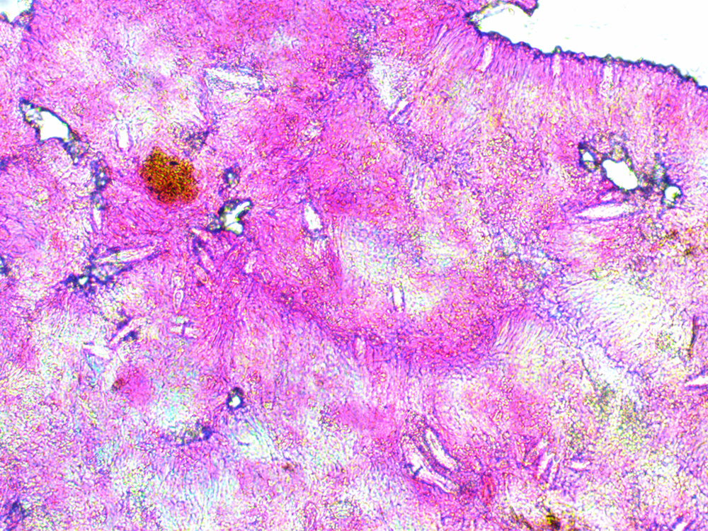
Glyeocystidia visible in top right of the image
and
then
enlarged in the image below.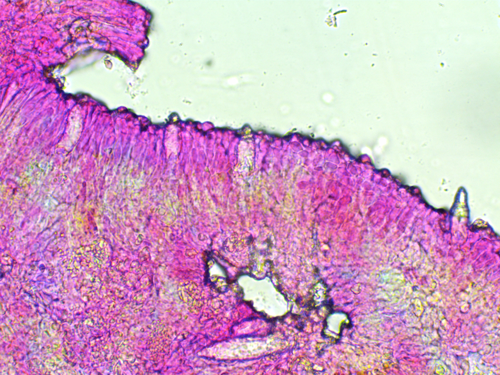
x100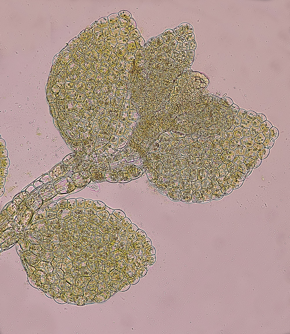
x200
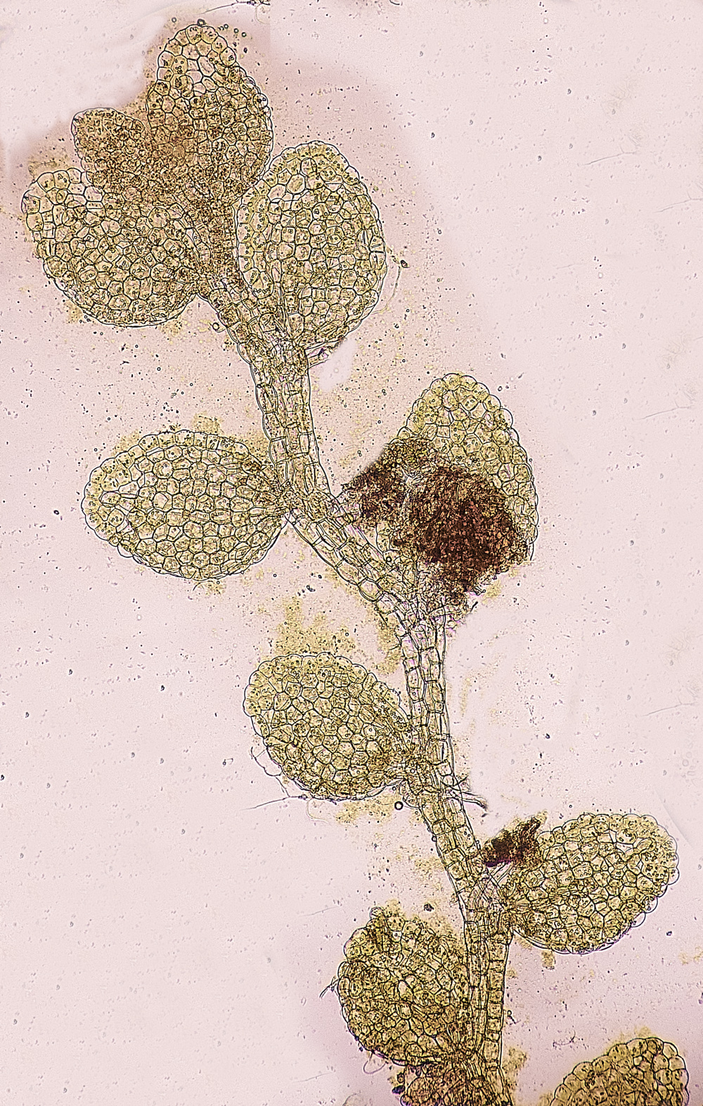
Minute Pouncewort (Cololejeunea minutissima)
This one was a lucky find as I've never seen it before. Tiny! 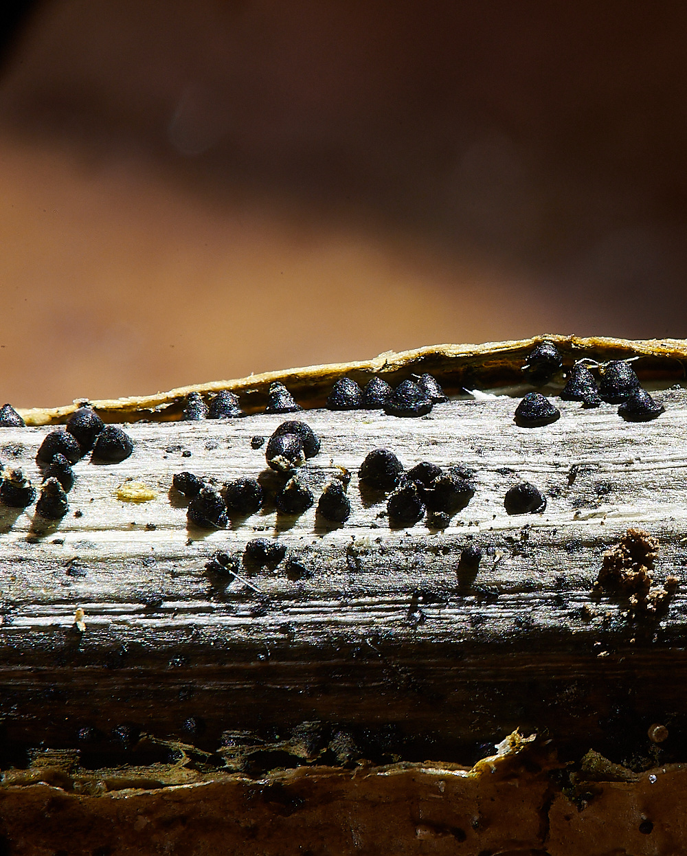
Nettle Rash (Leptosphaeria acuta)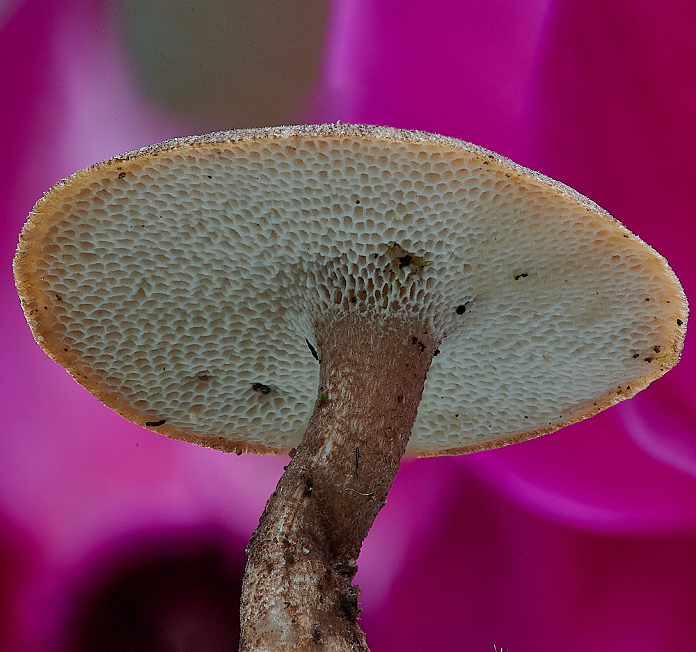
Winter Polypore (Polyporus brumalis)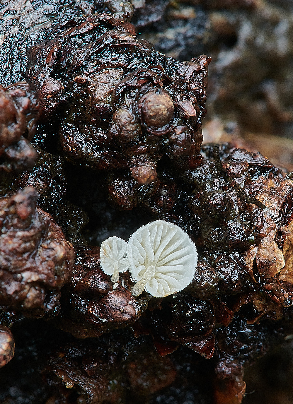
Dewdrop Bonnet (Hemimycena tortuosa)
From Yvonne
The tiny white one in the log which we thought might be a Clitopilus was in fact Hemimycena tortuosa - it had a grown a bit by the time I looked at it and looked more like a normal agaric, then I found the spiral pileocystidia on the cap ( by accident actually - trying to get a bit of gill ! )
Thank you for a great day.
